
Method and system for displaying medical images

Active Publication Date: 2007-06-07
GENERAL ELECTRIC CO
View PDF9 Cites 32 Cited by
  • Summary
  • Abstract
  • Description
  • Claims
  • Application Information

AI Technical Summary

Problems solved by technology

Analysis of the images can be very time sensitive or critical with added time possibly resulting in increased risk to the patient due to the patient condition.
This added time can result in increased risk of permanent injury or death.
Accordingly, rapidly determining the appropriate diagnosis and treatment path is often critical and can reduce the chances of permanent injury or death.
Increased time in diagnosis can increase the likelihood of permanent injury.
If the diagnosis is not correct, for example, if there is no stroke and a patient is given a stroke treatment, serious harm and death can result.
Improper diagnosis can lead to improper treatment.
Known methods for displaying medical image data to analyze for diagnosing a patient can be difficult to view or navigate, thereby increasing the time for diagnosis.
For example, due to the basic complexity of some anatomical regions, such as the brain, existing segmentation techniques do not allow for a clean visualization.
Similarly, these systems display a single image in time and may not provide the clinician with all of the information needed for a proper diagnosis.
The clinician often has to view different images, which may be on different screens or may not be able to be viewed simultaneously.
This increases the difficulty in reviewing and analyzing the images, thereby increasing the time for a proper diagnosis.
Thus, known methods and systems for displaying medical images can be difficult to navigate and review.
Further, these system can be cumbersome or awkward to use, further adding time before a proper diagnosis.
This added time can result in serious consequences.

Method used


Image

  • Method and system for displaying medical images
  • Method and system for displaying medical images
  • Method and system for displaying medical images

Examples


Embodiment Construction

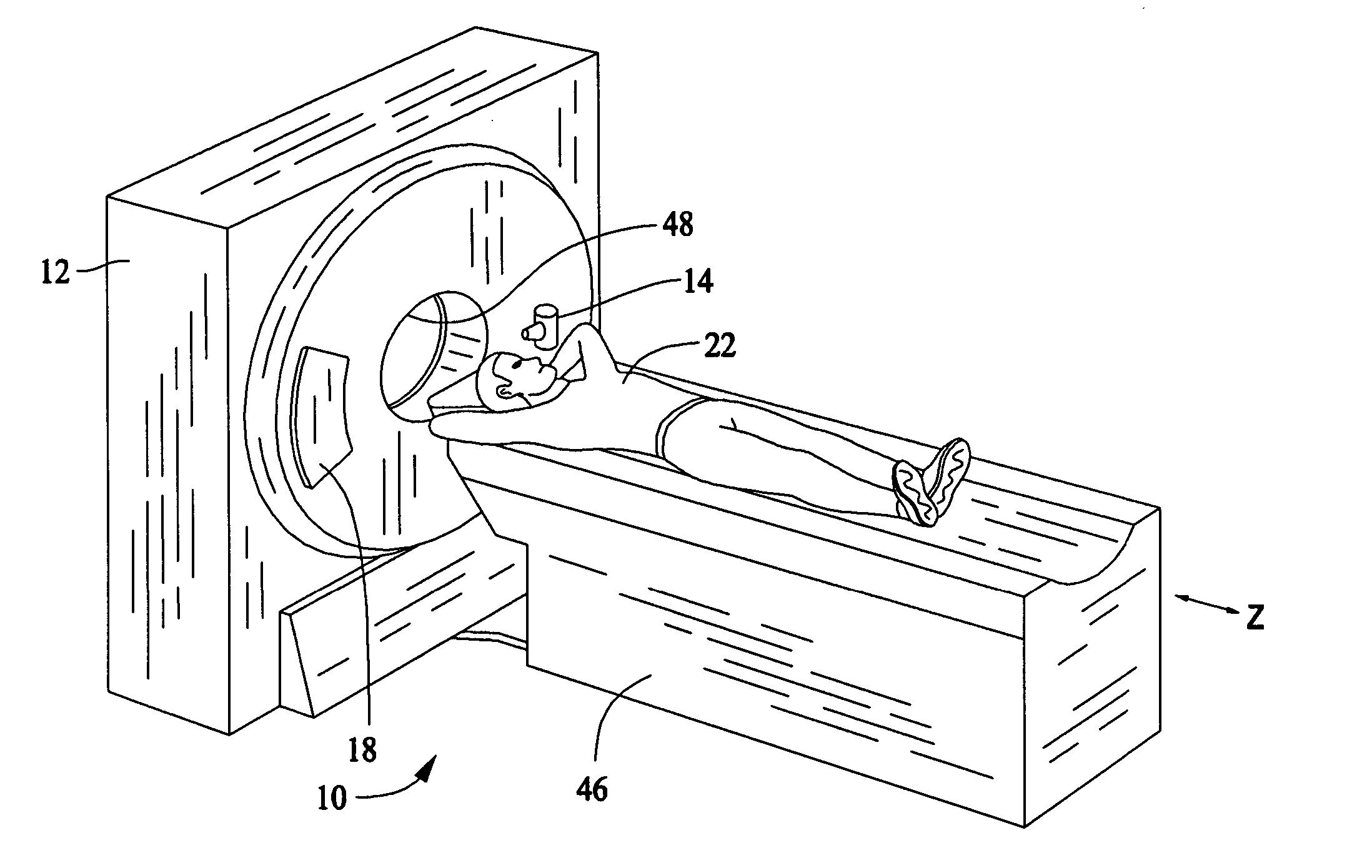
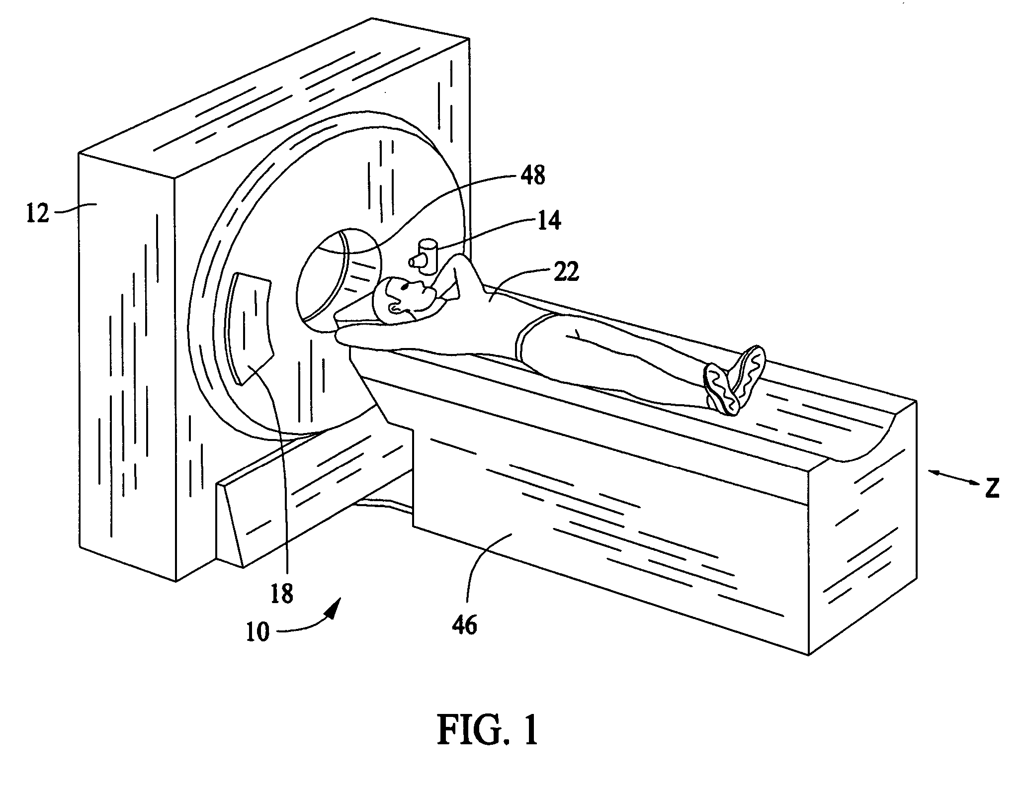
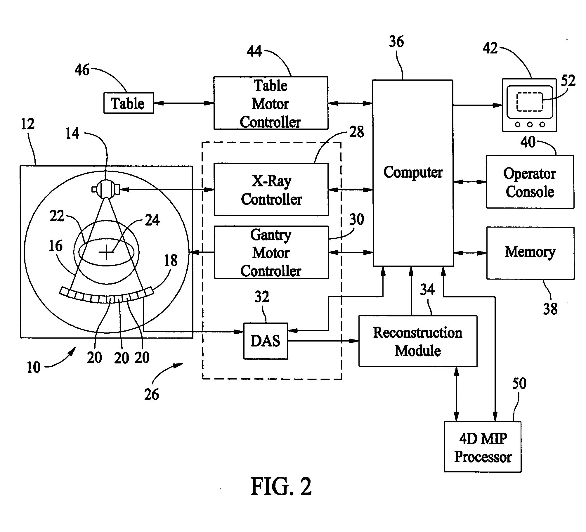
[0014]FIG. 1 is a perspective view of an exemplary imaging system 10. FIG. 2 is a schematic block diagram of the imaging system 10 (shown in FIG. 1). In an exemplary embodiment, the imaging system 10 is a single modality imaging system, for example, a computed tomography (CT) system. However, it should be understood that the various embodiments may be implemented in connection with imaging systems having more than one imaging modality (i.e., multi-modality imaging systems). Additionally, although the various embodiments may be described in connection with a particular imaging modality, for example, CT imaging, different imaging modalities both medical and non-medical may be used, for example, Positron Emission Tomography (PET), and in general any type of x-ray or nuclear imaging.

[0015] Referring now specifically to FIGS. 1 and 2, a computed tomography (CT) imaging system 10 is shown that includes a gantry 12 for a CT scanner. The gantry 12 includes an x-ray source 14 that projects ...


PUM


Abstract

Methods and systems for displaying medical images are provided. The method includes performing three-dimensional (3D) volume visualization on a plurality of two-dimensional (2D) images acquired continuously. The method further includes combining the 3D volume visualizations to form a plurality of four-dimensional (4D) volumes representing the 3D volume visualizations over a period of time and displaying at least one 4D volume.

Description

BACKGROUND OF THE INVENTION [0001] This invention relates generally to medical imaging systems, and more particularly, to systems that display images resulting from scans performed by medical imaging systems. [0002] Medical analysis and diagnosis is often performed with medical imaging system having displays to display the results of a scan of a patient. Analysis of the images can be very time sensitive or critical with added time possibly resulting in increased risk to the patient due to the patient condition. This added time can result in increased risk of permanent injury or death. For example, a brain attack is a time critical patient condition where every minute counts. Accordingly, rapidly determining the appropriate diagnosis and treatment path is often critical and can reduce the chances of permanent injury or death. In particular, for a patient showing signs of a possible stroke, it is important to quickly determine whether there is brain hemorrhaging. Increased time in dia...

Claims


Application Information

Patent Timeline
IPC IPC(8): A61B5/05A61B6/00
CPCA61B6/032A61B6/037A61B6/463A61B6/466A61B6/481A61B6/504
Inventor PROFIO, MARK VINCENTOKERLUND, DARINPROCKNOW, KARENHOLE, ANN CHRISTINE
Owner GENERAL ELECTRIC CO
Learn More