Method of inducing the differentiation of stem cells into myocardial cells

a stem cell and myocardial cell technology, applied in the field of stem cell differentiation into myocardial cells, can solve the problems of heart failure, cardiac function lowering, cardiomyocyte loss cannot be replaced, etc., and achieve the effect of efficient and selective production

Inactive Publication Date: 2007-06-14
FUKUDA
View PDF0 Cites 80 Cited by
  • Summary
  • Abstract
  • Description
  • Claims
  • Application Information

AI Technical Summary

Benefits of technology

[0087] Myocardial precursor cells and cardiomyocytes can be efficiently and selectively produced from stem cells using the method of the present invention. Myocardial (precursor) cells prepare

Problems solved by technology

In general, cardiomyocytes undergo active cell division with beating autonomously before birth, but immediately after birth they lose the ability to divide, and since they have little undifferentiated precursor cells, when cardiomyocytes die due to exposure to various forms of stress including myocardial infarction, myocarditis and the like, the lost cardiomyocytes cannot be replaced.
As a result, the surviving cardiomyocytes try to maintain myocardial function through compensatory hypertrophy and the like, but if the stress continues and exceeds an allowable threshold, it leads to further exhaustion and death of cardiomyocytes and a consequent lowering of myocardial function (that is, heart failure).
Heart failure and other types of heart disease are the second leading cause of death in Japan, and prognoses are very poor, with a 5-year survival rate of only about 50% for patients with heart diseases.
Conventional therapeutic drugs for heart failure include digitalis preparations that increase the contractive force of the myocardium and xanthine preparations and other heart stimulants, but long-term administration of these drugs is known to make the condition worse because there is too much expenditure of myocardial energy.
More recently, mainstream therapy has shifted to beta-blockers and ACE inhibitors, which reduce the excess burden on the heart due to s

Method used


Image

  • Method of inducing the differentiation of stem cells into myocardial cells
  • Method of inducing the differentiation of stem cells into myocardial cells
  • Method of inducing the differentiation of stem cells into myocardial cells

Examples


example 1

Effects of Noggin Treatment on Cardiomyocyte-Differentiation from ES Cells

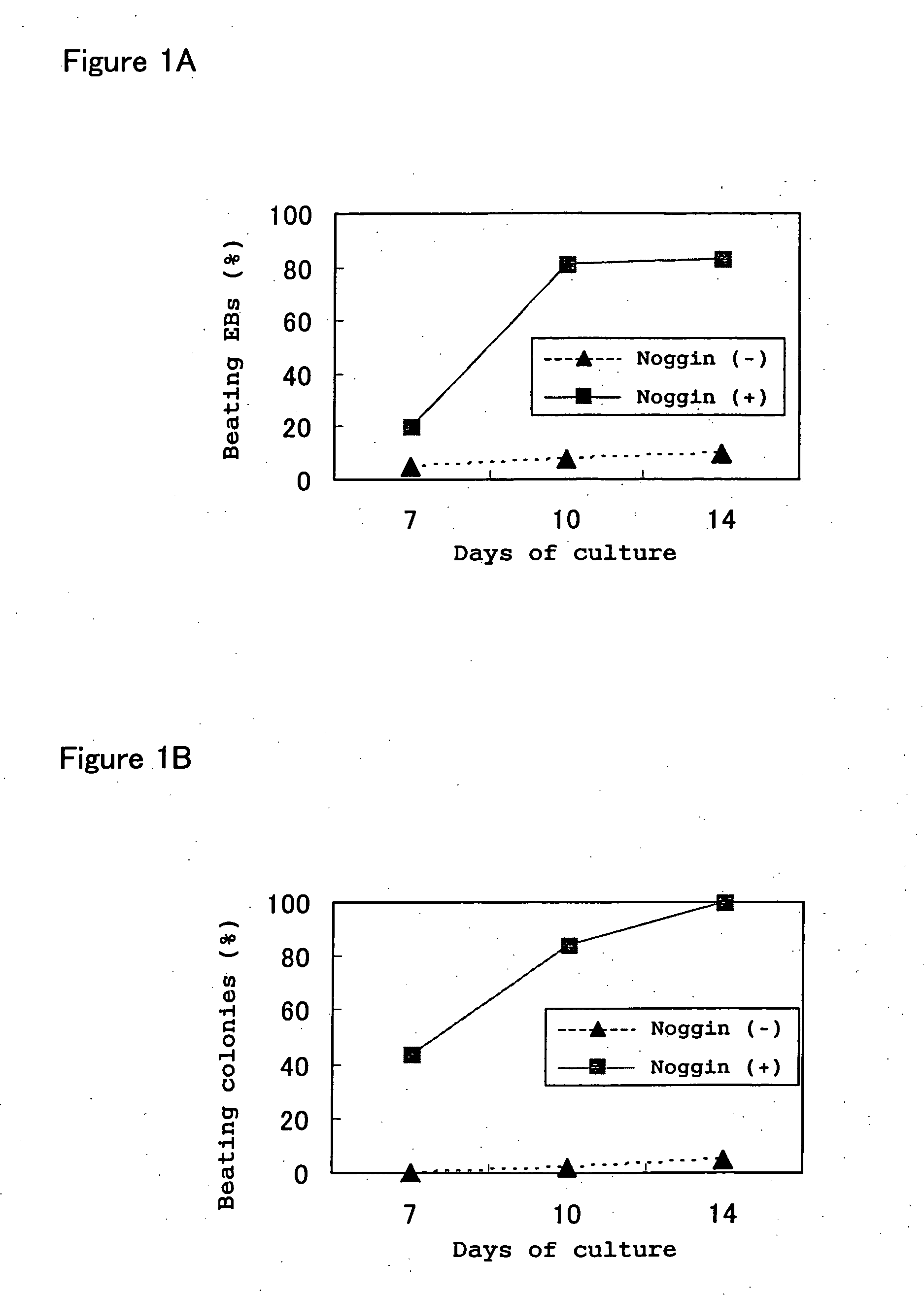
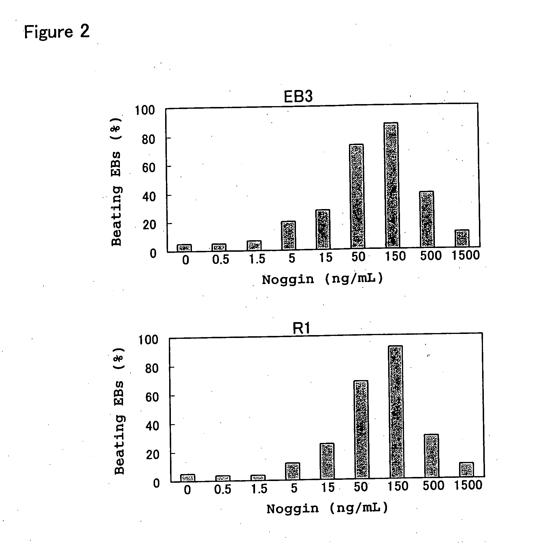
[0131] The effects of addition of the Noggin protein to culture medium on the development of cardiomyocytes were studied in an experimental system in which EBs were formed by floating aggregation culture (called simply floating culture in the examples of this application below) of ES cells and induced to differentiate into cardiomyocytes.

[0132] In the experiments below, EB3 cells (provided by Prof. Hitoshi Niwa of Riken, Japan), R1 cells (provided by Andrew Nagy of Mount Sinai Hospital, Canada) and 129SV cells (purchased from Dainippon Pharmaceutical Co.) were used as the ES cells, but in general there were no differences between these ES cell lines. These ES cells were passaged and maintained in an undifferentiated state according to the methods described in Manipulating the Mouse Embryo: A Laboratory Manual (Hogan et al Eds., Cold. Spring Harbor Laboratory Press, 1994), Embryonic Stem Cells: Methods and Pr...

example 2

Properties of Cardiomyocytes Derived from Noggin-Treated ES Cells

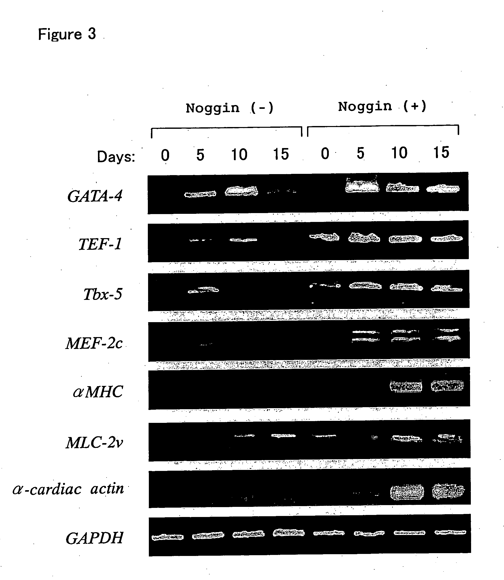
[0140] As shown in Example 1, beating of EBs prepared from ES cells was increased significantly by Noggin treatment, and to confirm that the beating cells in these EBs were cardiomyocytes, we investigated the expression of various myocardial-specific marker genes and proteins.

[0141] Expression of various myocardial cell-specific marker genes in the Noggin (+) group and Noggin (−) group is shown in FIG. 3. EBs formed by floating culture as in FIG. 1 were collected periodically, and total RNA was prepared using RNeasy (Qiagen). Next, cDNA was reverse-transcribed by conventional methods using Superscript II (Invitrogen), and genes specific to cardiomyocytes were detected by polymerase chain reaction (PCR). The primers used to detect the transcripts of GATA-4, TEF-1, Tbx-5, MEF-2c, αMHC, MLC-2v, α-cardiac actin and GAPDH were as follows.

GATA-4 (forward)5′-CTGTCATCTC ACTATGGGCA-3′(SEQ ID NO:1)GATA-4 (reverse)5′-CCAAGTCC...

example 3

Effects of Differences in Noggin Treatment Time and Period on Cardiomyocyte Differentiation from ES Cells

[0148] To determine the optimum time and period for Noggin treatment, the time of addition of the Noggin protein to the aforementioned floating culture system was varied, and the subsequent effects on myocardial differentiation induction were examined.

[0149] The results are shown in Table 1.

TABLE 1Pre-differentiationDifferentiation-stageinducing stageAppearance ofNogginNogginBeating EBs−−++ 80%−++− 

[0150] Table 1 shows the appearance rates of beating EBs (EB3 cell derived) under various culture protocols, indicating the importance of Noggin stimulus before and after EBs formation. In Table 1, “pre-differentiation stage” indicates culture conditions from 3 days before until the start of floating culture, while “differentiation-inducing stage” indicates culture conditions at the point of EBs formation. In Table 1, “+” indicates than Noggin (150 ng / mL) was added to the medium an...


PUM

PropertyMeasurementUnit
Timeaaaaaaaaaa
Timeaaaaaaaaaa
Morphologyaaaaaaaaaa

Abstract

For a method of inducing differentiation of cardiomyocytes from stem cells, a method is provided to induce efficiently and selectively differentiation of cardiomyocytes by such a method in which the stem cells are cultured to induce differentiation into cardiomyocytes in the presence of a substance that inhibits BMP signaling.

Description

TECHNICAL FIELD [0001] The present invention relates to a method of preparing cardiomyocytes selectively and efficiently from ES cells and other pluripotent stem cells, and to cells for regenerative medicine obtained by this method. BACKGROUND ART [0002] In general, cardiomyocytes undergo active cell division with beating autonomously before birth, but immediately after birth they lose the ability to divide, and since they have little undifferentiated precursor cells, when cardiomyocytes die due to exposure to various forms of stress including myocardial infarction, myocarditis and the like, the lost cardiomyocytes cannot be replaced. As a result, the surviving cardiomyocytes try to maintain myocardial function through compensatory hypertrophy and the like, but if the stress continues and exceeds an allowable threshold, it leads to further exhaustion and death of cardiomyocytes and a consequent lowering of myocardial function (that is, heart failure). [0003] Heart failure and other ...

Claims


Application Information

Patent Timeline
IPC IPC(8): A61K48/00C12N5/08C12N5/077A61K35/12A61K35/545A61P9/04C12N5/07C12N5/071C12N5/0735C12N5/074C12N5/0789C12N15/09
CPCC12N5/0657C12N2501/155C12N2506/03A61P9/04C12N5/06C12N5/0652
Inventor FUKUDAYUASA, SHINSUKEOKANO, HIDEYUKISHIMAZAKI, TAKUYAKOSHIMIZU, UICHITANAKA, TOMOFUMISUGIMURA, KEIJIRO
Owner FUKUDA
Why Eureka
Try Eureka