Imaging catheter and method for volumetric ultrasound

a volumetric ultrasound and imaging catheter technology, applied in the field of imaging catheters, can solve the problems of preventing the acquisition of high-quality real-time three-dimensional (rt3d) volumetric images, generally referred to as volumes, and the number of signal conductors that can physically fit within the limited size of the catheter, and the limitation is particularly severe for two-dimensional arrays

Inactive Publication Date: 2007-07-19
GENERAL ELECTRIC CO
View PDF13 Cites 32 Cited by
  • Summary
  • Abstract
  • Description
  • Claims
  • Application Information

AI Technical Summary

Problems solved by technology

For severely space-constrained applications, such as imaging from catheter-mounted transducers (intracardiac echocardiography or ICE), two major limitations prevent the acquisition of high quality real-time three-dimensional (RT3D) volumetric images, generally referred to as volumes.
The first limitation is due to the number of signal conductors that can physically fit within the limited size of the catheter.
This limitation is especially severe for two-dimensional arrays, which can be electronically scanned in two dimensions to form RT3D volumes, because they typically require M×N connections, where M and N are the number of rows and columns of transducer elements in the array, respectively.
The second limitation is due to the small physical size available for the acoustic aperture.
Transducers confined to this spatial extent typically generate ultrasound beams that diverge rapidly with distance, resulti

Method used


Image

  • Imaging catheter and method for volumetric ultrasound
  • Imaging catheter and method for volumetric ultrasound
  • Imaging catheter and method for volumetric ultrasound

Examples


Embodiment Construction

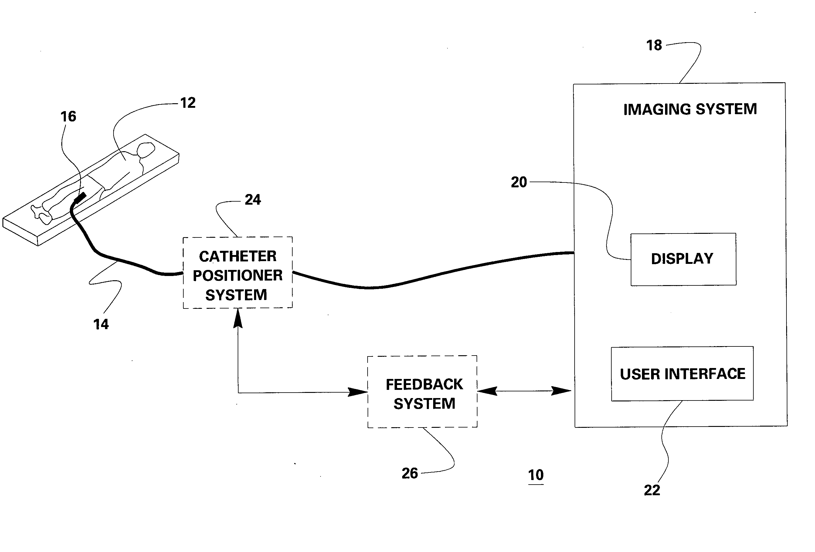
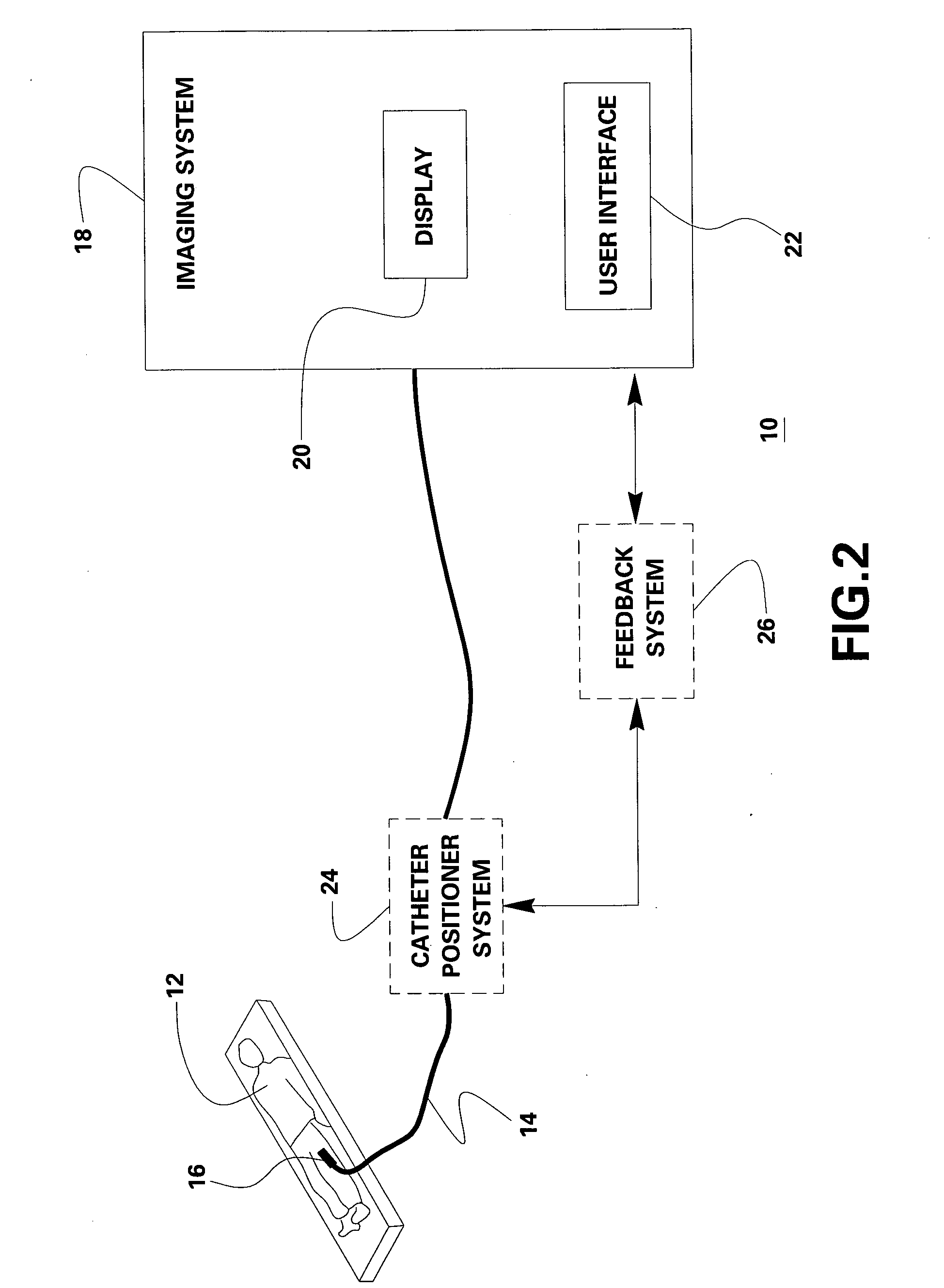
[0017] As will be described in detail hereinafter, a catheter assembly in accordance with exemplary aspects of the present technique is presented. Based on image data acquired by the catheter assembly, a three-dimensional volume of an anatomical region may be imaged and diagnostic information and / or the need for therapy in the anatomical region may be obtained.

[0018] In accordance with aspects of the present invention, the aforementioned limitations are overcome by using a transducer array that acquires image data at a given image plane and the transducer array is translated mechanically, or the active portion of the transducer is array is translated electronically, in a direction perpendicular to the image plane in order to image a 3D volume. The elements of the transducer array are electronically phased in order to acquire a sector image perpendicular to the long axis of the catheter, and the array is translated along the catheter axis in order to acquire the three-dimensional vo...


PUM


Abstract

An imaging catheter assembly and method for use in volumetric ultrasound imaging and catheter-guided procedures are provided. The imaging catheter assembly comprises a transducer array for acquiring image data at a given image plane and a motion controller coupled to the transducer array for translating the transducer array along a direction perpendicular to a direction of the image plane in order to image a three-dimensional (3D) volume.

Description

BACKGROUND [0001] The invention relates generally to an imaging catheter, and more particularly to a transducer array assembly for use in volumetric ultrasound imaging and catheter-guided treatment such as cardiac interventional procedures. [0002] In ultrasound imaging, spatial resolution is a critical factor in image quality. For severely space-constrained applications, such as imaging from catheter-mounted transducers (intracardiac echocardiography or ICE), two major limitations prevent the acquisition of high quality real-time three-dimensional (RT3D) volumetric images, generally referred to as volumes. The first limitation is due to the number of signal conductors that can physically fit within the limited size of the catheter. This limitation is especially severe for two-dimensional arrays, which can be electronically scanned in two dimensions to form RT3D volumes, because they typically require M×N connections, where M and N are the number of rows and columns of transducer ele...

Claims


Application Information

Patent Timeline
IPC IPC(8): A61B8/14
CPCA61B8/12A61B8/14A61B8/445A61B8/483A61B8/4461
Inventor LEE, WARRENRIGBY, KENNETH WAYNE
Owner GENERAL ELECTRIC CO
Why Eureka
Try Eureka