Method for non-cooperative lung function diagnosis using ultrasound
a non-cooperative, ultrasound technology, applied in the field of lung function diagnosis devices and methods, can solve the problems of only being able to perform the test successfully, the patient's exhaustion, and the test is relatively difficult to explain, and achieve the effect of simple use and low cos
- Summary
- Abstract
- Description
- Claims
- Application Information
AI Technical Summary
Problems solved by technology
Method used
Image
Examples
example 1
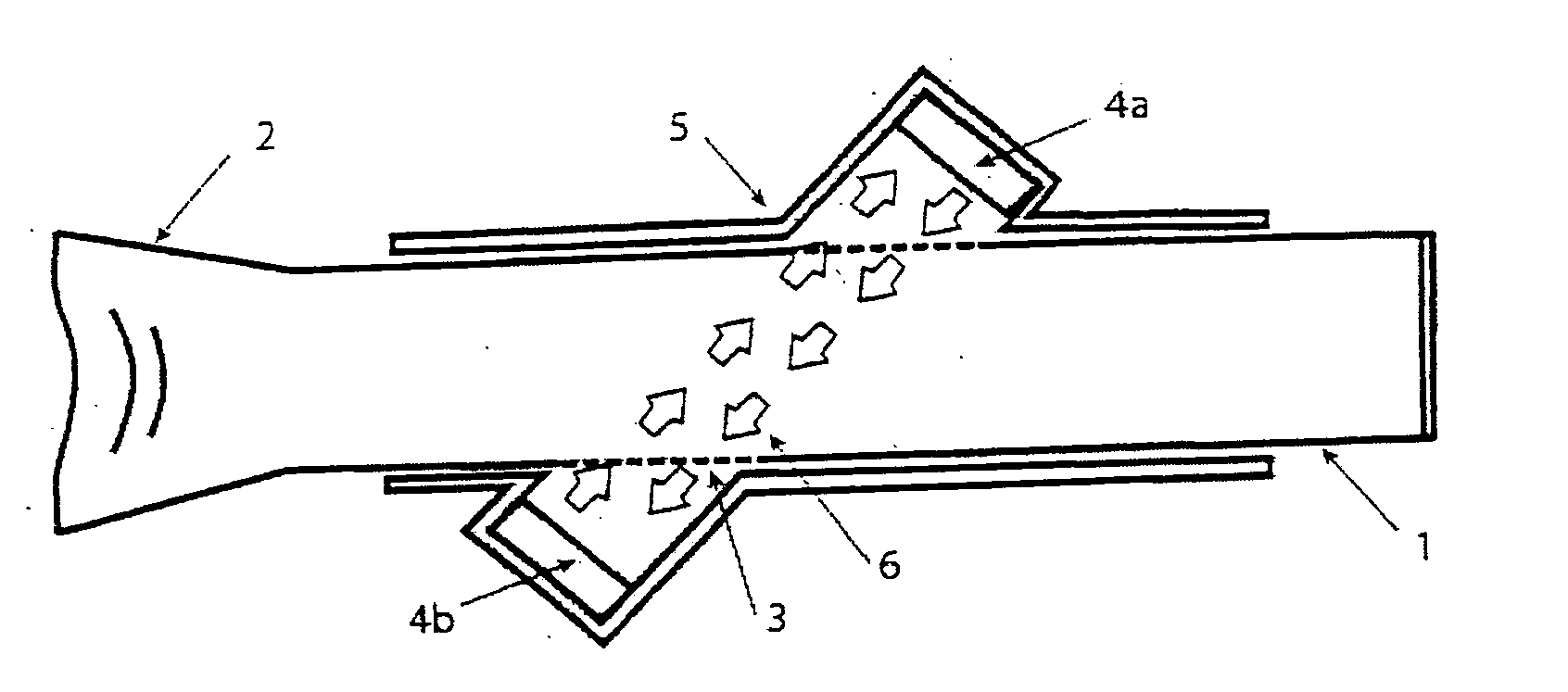
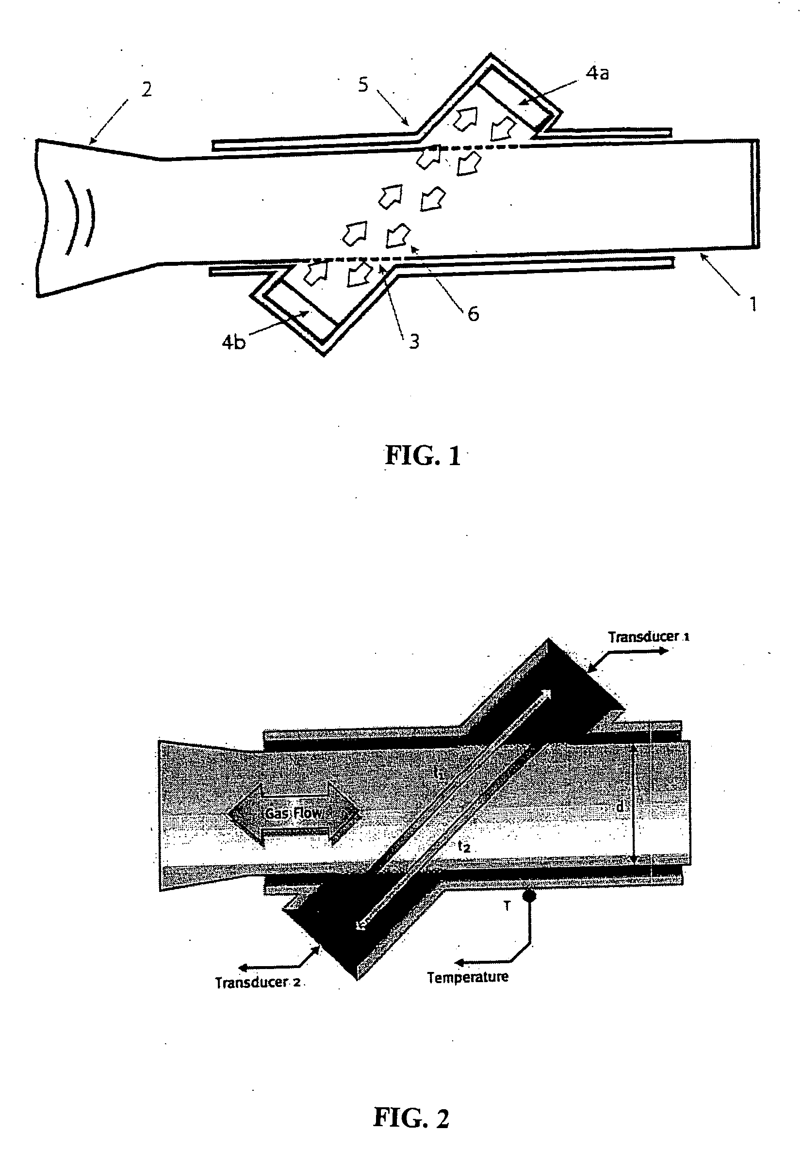
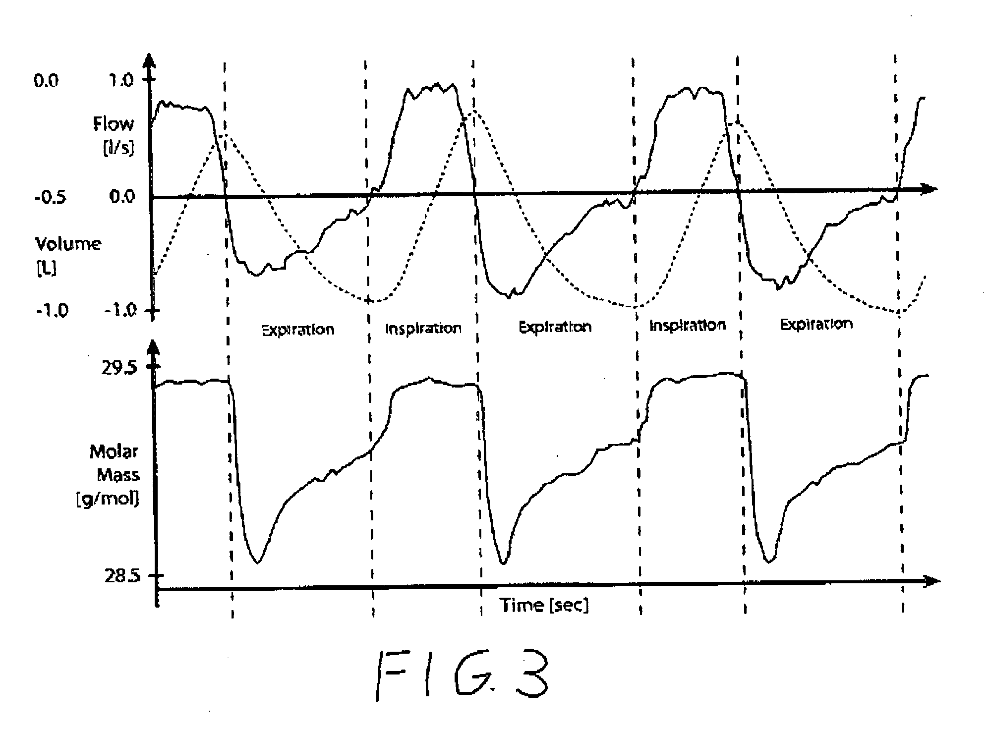
[0039] Patients with airway obstruction can be identified and categorized as to severity with measures of CO2 and spirometric flow patterns during quiet breathing. Sixty five healthy subjects and patients (35 men, 30 women) with mean age of 48.4 were tested in the pulmonary laboratory. They breathed quietly for 5 minutes on an ndd ultrasonic spirometer (ndd, Zurich, Switzerland) while seated using nose clips. Standard spirometry was conducted on all subjects, except one, prior to performing the quiet breathing on the ultrasonic molar mass device. PFT measurements of FVC and FEV1 and percent predicted values (using NHANES III reference equations) were obtained and used to categorize the subjects into normal, mild, moderate or severe obstruction or restricted. Diffusion capacity (DLCO) was measured only in scheduled patients.
[0040] Molar Mass (MM) measurement is a potential surrogate for CO2 measurement in pulmonary function testing. Molar mass measurements were compared to CO2 measu...
example 2
[0050] Patients with airway obstruction can be identified and categorized as to severity with measures of CO2 and spirometric flow patterns during quiet breathing. Molar mass measures were compared to CO2 measurements made with a mass spectrometer. Thirty six individuals were tested in the pulmonary laboratory. Clinical classifications from spirometry, DLCO and physician diagnosis is carried out on sixteen healthy subjects, four patients with mild obstruction, six patients with moderate obstruction and ten patients with severe obstruction.
[0051] Average values for FEV1 and FEV1 / FVC in each disease category are listed in Table 4, along with mean parameter values. There is excellent correlation between the parameters (1-4) derived from the mass spectrometer measured % CO2 vs. volume curves and the ultrasonic measured molar mass vs. volume curves.
TABLE 4NormalMildModerateSevereFEV1 (L)3.11 ± 0.753.24 ± 0.841.91 ± 0.741.55 ± 0.55FVC / FEV1 (%)77.9 ± 8.6 66.3 ± 3.0 59.3 ± 6.6 46.4 ± 8.9...
PUM
 Abstract
Description
Claims
Application Information
 - R&D
- Intellectual Property
- Life Sciences
- Materials
- Tech Scout
- Unparalleled Data Quality
- Higher Quality Content
- 60% Fewer Hallucinations
Browse by: Latest US Patents, China's latest patents, Technical Efficacy Thesaurus, Application Domain, Technology Topic, Popular Technical Reports.
© 2025 PatSnap. All rights reserved.Legal|Privacy policy|Modern Slavery Act Transparency Statement|Sitemap|About US| Contact US: help@patsnap.com


