
Virtual Surgical System and Methods

a virtual surgery and computer system technology, applied in the field of virtual surgery implementations of computer systems, can solve the problems of bone fragment malalignment, bone fragments that are difficult to restore the form and function of craniofacial skeleton, and require a degree of precision

Inactive Publication Date: 2007-09-06
UNIV OF GEORGIA RES FOUND INC
View PDF13 Cites 20 Cited by
  • Summary
  • Abstract
  • Description
  • Claims
  • Application Information

AI Technical Summary

Problems solved by technology

Although any alignment of bone fragments requires a degree of precision, restoring the form and function of the bone fragments in the craniofacial skeleton is particularly challenging because of the strict tolerances required.
There are several problems with this current, standard approach.
However, with decreased dissection, the surgeon not being able to visualize the entire fracture, which could lead to potential mal-alignments of the bone fragments.
Further, because the surgeon does not determine the type and configuration of the prosthesis to use until the surgery is underway, the surgery is prolonged while the prostheses is configured or manufactured appropriately.
This results in extended periods of time in which the patient remains dissected, increasing the chance of surgical complications and tying up valuable surgical resources such as surgical clean rooms, surgeons, nurses, and anesthesiologists, etc.

Method used


Image

  • Virtual Surgical System and Methods
  • Virtual Surgical System and Methods
  • Virtual Surgical System and Methods

Examples


embodiment 300

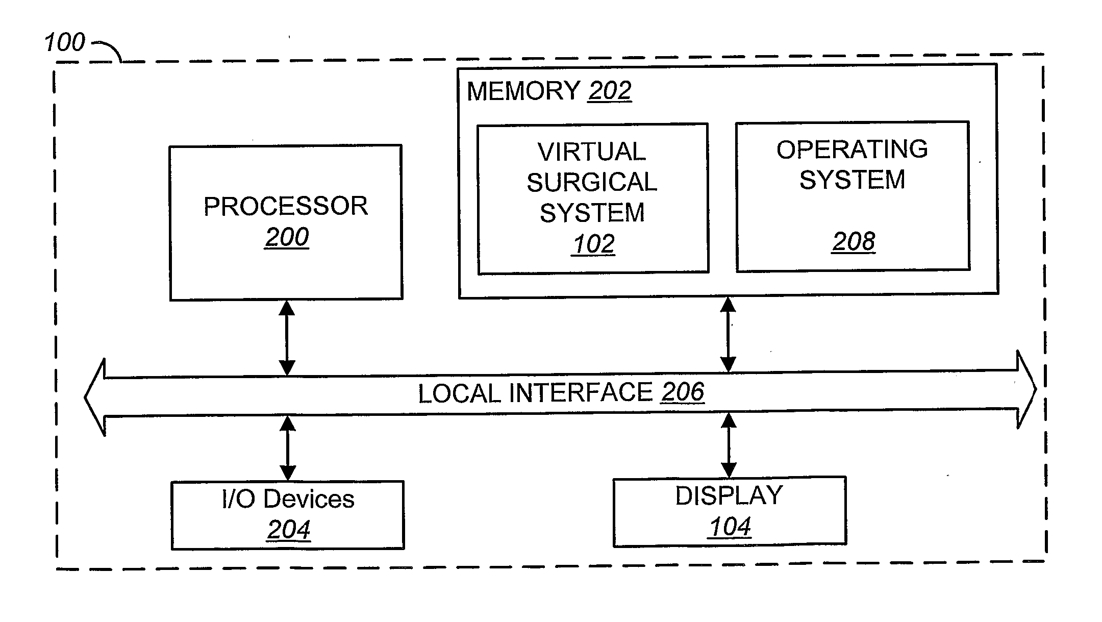
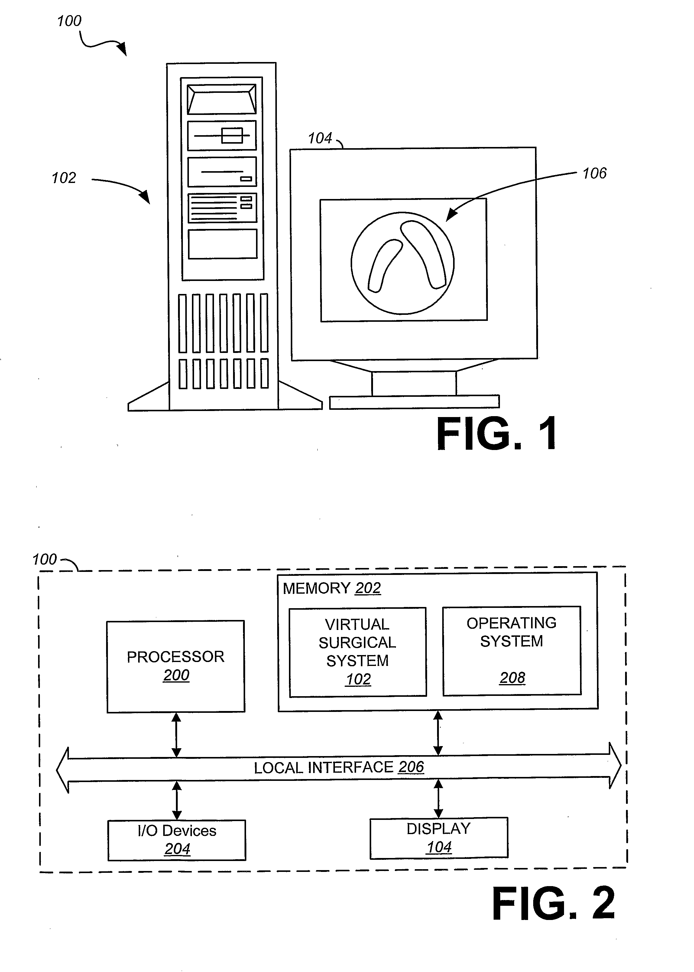
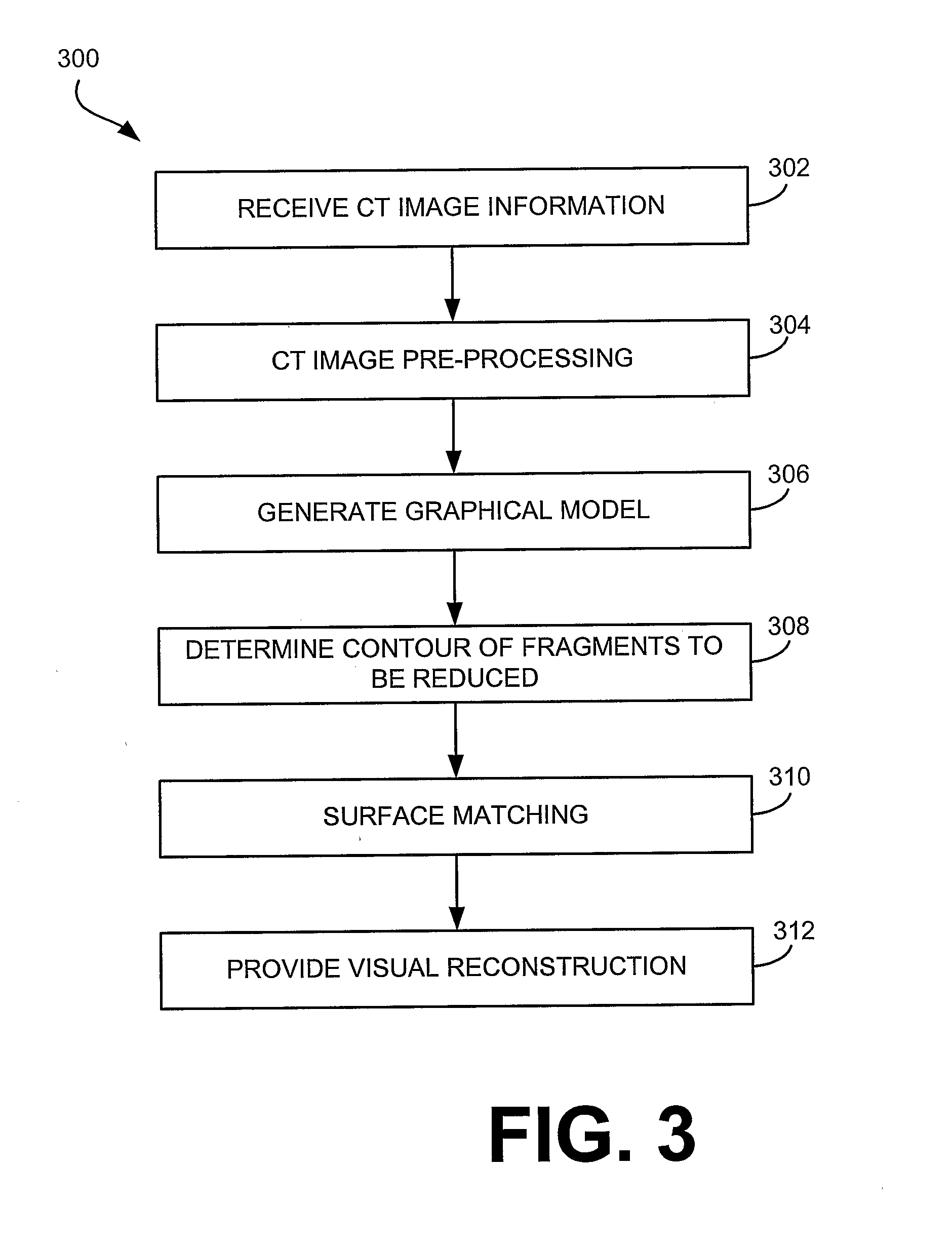
[0043] As depicted in the method embodiment 300, step 302 includes receiving information representing at least one digital image of fragmented bone elements. For example, one input of digital image information to the virtual surgical system 102 may be a sequence of grayscale images of a fractured human mandible. In the present embodiment, the images are two-dimensional (2-D) computer tomography (CT) image slices. In one embodiment, exemplary dimensions and color features for the CT image slices may be 150 millimeter (mm)×150 mm with an 8-bit color depth. The digital image information may be provided, for example, across local interface 206 from input devices 204.

[0044] Step 304 includes pre-processing the digital image information (eg. the CT image). Specifically, as depicted in FIG. 4, in one embodiment, a series of image pre-processing tasks 304a-304b are undertaken by the virtual surgical system 102 before using the surface matching algorithms to obtain the desired goal of reduct...

embodiment 500

[0055]FIG. 5 depicts a user-assisted, interactive contour detection embodiment 500, which may be performed to determine the contour as illustrated in step 308 of FIG. 3. At step 308a, a user is iteratively guided through one or more graphical interfaces to select (e.g. via a mouse, stylus, or other input means) end points of a fracture contour in the CT slices. Exemplary user interfaces for the interactive contour detection are described in more detail below with respect to FIGS. 12-14. However, the contour points may be selected, for example, upon the user selection (e.g. a mouse button click, key press, etc.) of a particular area (e.g. a pixel, or group of pixels) according to the position of a cursor (e.g. guided by a mouse or other input device) over the two-dimensional CT scan image. The user performing the selection may, for example, be a surgeon or one skilled in performing such operations. The virtual surgical system 102 collects the data corresponding to the position of the...


PUM


Abstract

A virtual surgical system receives an image, such as a computer tomography (CT) scan, of an area of the patient in need of surgical reconstruction prior to an incision being made in a surgery. Bone fragments that are to be reduced are automatically selected using the virtual surgical system. The system performs surface matching techniques using surface data representing the contour of the end surfaces of the fragments to determine how the corresponding fragments should be oriented to perform the reduction. At least one of the fragments may then be transformed (e.g. via translation and / or rotation) to join the surfaces based on the matched surfaces. An image may be generated of the reconstructed area providing a virtual representation of a reconstructed object. Thus, before actual dissection of the area, and the subsequent physical surgical reduction of the bones, an image of the reconstructed object may be displayed such that a prosthesis may be determined and / or manufactured.

Description

CROSS-REFERENCE TO RELATED APPLICATIONS [0001] This application claims priority to, and the benefit of, U.S. Provisional Patent Application entitled “Virtual Surgical Systems and Methods,” assigned Ser. No. 60 / 561,584, and filed on Apr. 13, 2004, which is incorporated by reference in its entirety.TECHNICAL FIELD [0002] The present disclosure is generally related to computer systems and, more particularly, is related to computer systems for virtual surgery implementations. BACKGROUND [0003] Traumatic impact of the skeleton may require extensive surgery to re-align fragmented skeletal features. Although any alignment of bone fragments requires a degree of precision, restoring the form and function of the bone fragments in the craniofacial skeleton is particularly challenging because of the strict tolerances required. During conventional craniofacial surgery, a plastic surgeon restores the form and function of the bone fragments in the craniofacial skeleton by first exposing all the fr...

Claims


Application Information

Patent Timeline
IPC IPC(8): A61B5/00A61B19/00G16H20/40G16H30/40G16H40/67G16Z99/00
CPCA61B19/2203A61B19/50A61B19/52A61B19/56A61B2019/5236G06T2207/30008G06F19/3418G06F19/3437G06T7/0012G06T2207/10081A61B2019/5238A61B90/36A61B34/25A61B2090/376A61B34/30A61B34/10A61B2090/374G16H50/50G16H20/40G16H30/40G16H40/67G16Z99/00
Inventor BHANDARKAR, SUCHENDRA M.TOLLNER, ERNEST WILLIAM
Owner UNIV OF GEORGIA RES FOUND INC
Learn More