Method for imaging an infarction patient's myocardium and method for supporting a therapeutic intervention on the heart

a technology for infarction patients and myocardium, which is applied in the field of imaging the myocardium of infarction patients and the method of supporting a therapeutic intervention on the heart, can solve the problems of not being able to treat necrotic myocardial tissue, not being able to gain information during an intervention about the patient, and damage to the myocardium (heart tissue)

Inactive Publication Date: 2007-10-04
SIEMENS AG
View PDF9 Cites 7 Cited by
  • Summary
  • Abstract
  • Description
  • Claims
  • Application Information

AI Technical Summary

Benefits of technology

[0034]The detailed image data further permits the following additional invention: The coordinates of a boundary between the necrotic myocardium and other areas of tissue are determined using the image representations produced at step o) (or, if o) is omitted, using the superimposed image data obtained at step n)). Registering having, of course, taken place at step m), that is done in the patient system used during the intervention, even if the necrotic myocardium has been pre-operatively recorded. That will make it possible for a therapy instrument (for example an ablation catheter) to be moved to at least one point on the boundary under automatic control (by means of a suitable control motor). The ablation catheter can preferably even be moved along the entire boundary. As mentioned in the introduction, the boundary between the necrotic myocardium and the parts thereof having a reduced blood supply is of interest particularly for ablating because, on the one hand, ablating aimed at restoring the blood supply to the necrotic myocardium will serve little purpose as that is by definition already dead; on the other hand, ablating specifically at the boundary between necrotic myocardium and myocardium having a reduced blood supply will be to particularly good purpose. The inventive method can thus be advanced to the extent that the doctor can let the system operate autonomously and will only have to intervene supportively.

Problems solved by technology

The myocardium (heart tissue) can be damaged during a myocardial infarction.
It is not possible to treat necrotic myocardial tissue.
No information can be gained during an intervention about whether further coronary arteries or further sub-branches of the artery are to be treated.
There is no supporting imaging method that provides a representation in which necrotic tissue and tissue having a reduced blood supply are imaged simultaneously adequately well.
However, with none of said imaging methods is tissue having a reduced blood supply visualized adequately well alongside the necrotic myocardial tissue.

Method used


Image

  • Method for imaging an infarction patient's myocardium and method for supporting a therapeutic intervention on the heart
  • Method for imaging an infarction patient's myocardium and method for supporting a therapeutic intervention on the heart
  • Method for imaging an infarction patient's myocardium and method for supporting a therapeutic intervention on the heart

Examples


Embodiment Construction

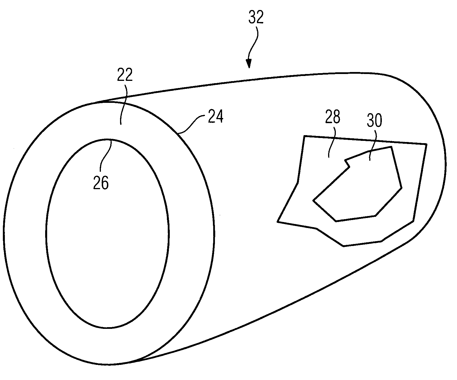
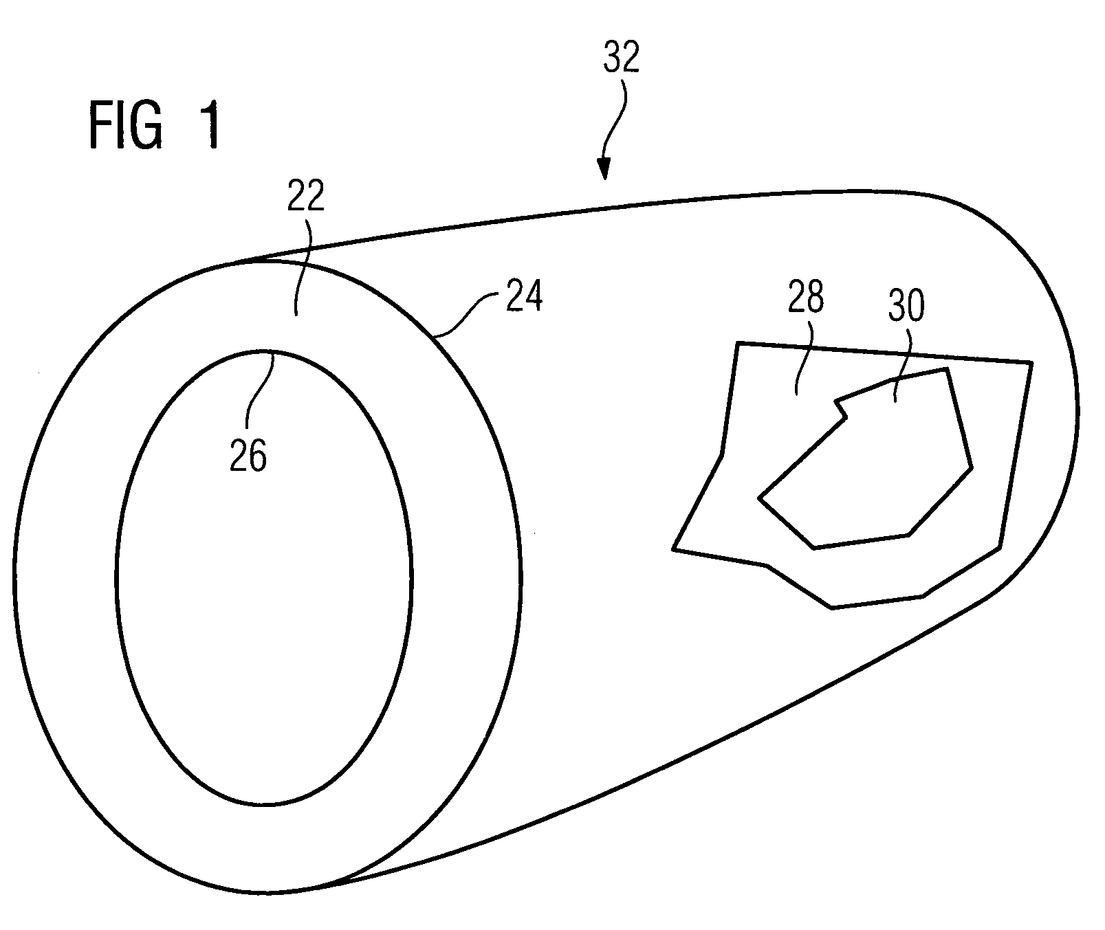
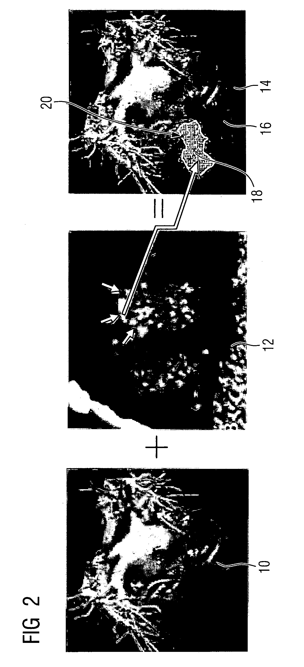
[0047]In the inventive method a 3D image data record is first produced in which can be seen endocardium, healthy parts of the myocardium, and parts thereof having a reduced blood supply.

[0048]A contrast medium is therein used in the conventional manner. An image representation of said kind is known from the prior art.

[0049]It is further known from the prior art cited in the introduction how to produce a second 3D image data record in which the necrotic myocardial regions are particularly well imaged. Although a contrast medium is employed for that purpose, a period of time is allowed to elapse until said medium has dispersed. As the contrast medium collects mainly in the necrotic parts of the myocardium, those are particularly well imaged.

[0050]Two 3D image data records are hence available. These are then to be used for producing a superimposed representation. The first 3D image data record produced is for that purpose segmented. The method of segmenting is as such known in the prio...


PUM


Abstract

Two 3D image data records are obtained mutually independently comprising healthy myocardium, myocardium having a reduced blood supply, and the necrotic myocardium. The image data records are combined to produce an overall image data record after registration, and 2D image representations are produced from the overall image data record in which the necrotic parts of the myocardium are shown emphasized, with simultaneously showing the endocardium or the healthy parts of the myocardium and parts having a reduced blood supply. The overall image data record can be used afterwards. For example, further registering step is carried out using images obtained during an intervention on the myocardium. The further registering step enables the necrotic parts of the myocardium to be assigned to the patient's situation. That can extend as far as catheters being moved automatically up to a boundary of the necrotic myocardium.

Description

CROSS REFERENCE TO RELATED APPLICATIONS[0001]This application claims priority of German application No. 10 2006 014 882.7 filed Mar. 30, 2006, which is incorporated by reference herein in its entirety.FIELD OF THE INVENTION[0002]The invention relates to a method for imaging an infarction patient's myocardium. It relates also to a method for supporting a therapeutic intervention on the heart.BACKGROUND OF THE INVENTION[0003]The myocardium (heart tissue) can be damaged during a myocardial infarction. A distinction is made following a myocardial infarction between healthy myocardium being supplied normally with blood, myocardium that has a reduced blood supply (but is not yet necrotic), and necrotic myocardium that is dead.[0004]Cardiological interventions or, later, also electrophysiological procedures are frequently carried out following an acute myocardial infarction in order to treat the patient. It is not possible to treat necrotic myocardial tissue. The starting point is as a rul...

Claims


Application Information

Patent Timeline
Patent Type & Authority Applications(United States)
IPC IPC(8): A61B5/05
CPCA61B5/0402A61B6/4441A61B6/463A61B6/466A61B6/481A61B6/503G06T2207/30101A61B6/541A61B18/1492A61B2018/00577G06T7/0012G06T7/0024A61B6/504G06T7/30A61B5/318
Inventor BOESE, JANRAHN, NORBERTLAUTENSCHLAGER, STEFAN
Owner SIEMENS AG
Why Eureka
Try Eureka