
Endoscopic Translumenal Surgical Systems

a technology of endoscopy and lumen, which is applied in the field of endoscopy translumenal surgery, can solve the problems of difficult positioning of the opening and re-inserting the endoscope, and the size and shape of the instruments that may be passed through the bodily lumen in order to perform a medical procedure in the abdominal cavity

Inactive Publication Date: 2007-11-08
ETHICON ENDO SURGERY INC
View PDF79 Cites 161 Cited by
  • Summary
  • Abstract
  • Description
  • Claims
  • Application Information

AI Technical Summary

Benefits of technology

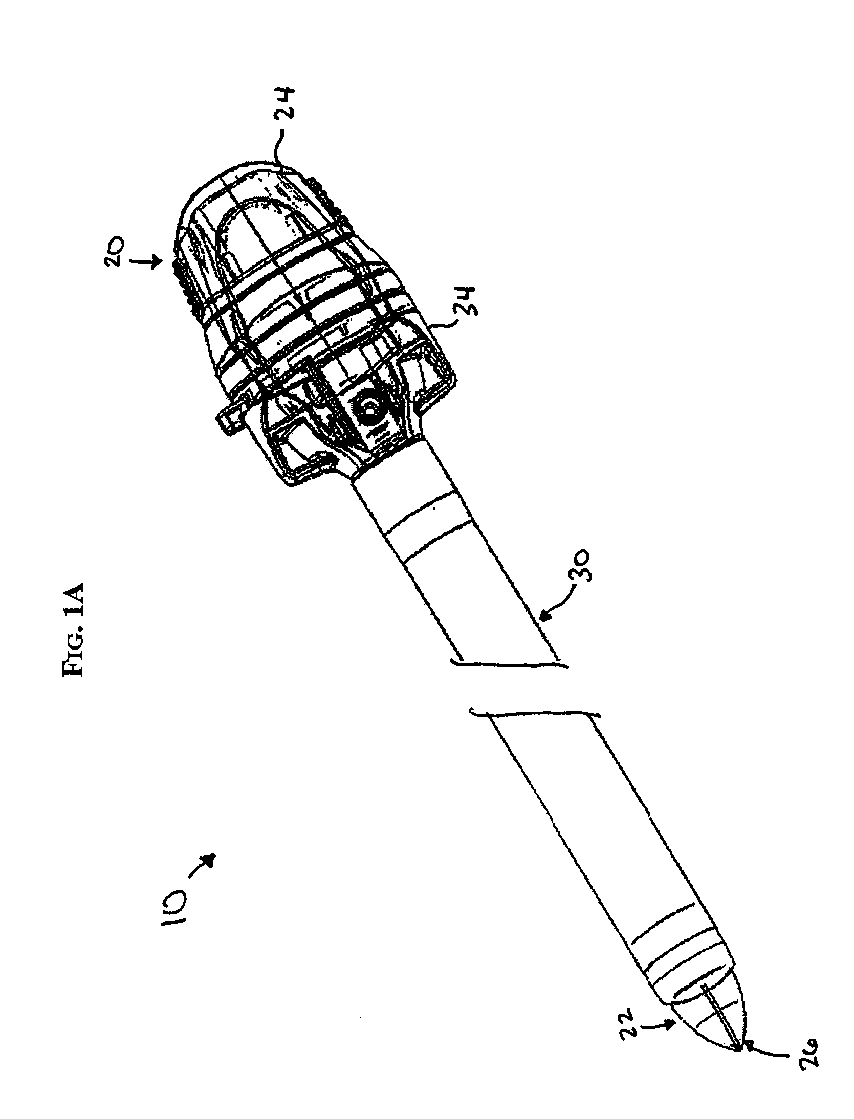
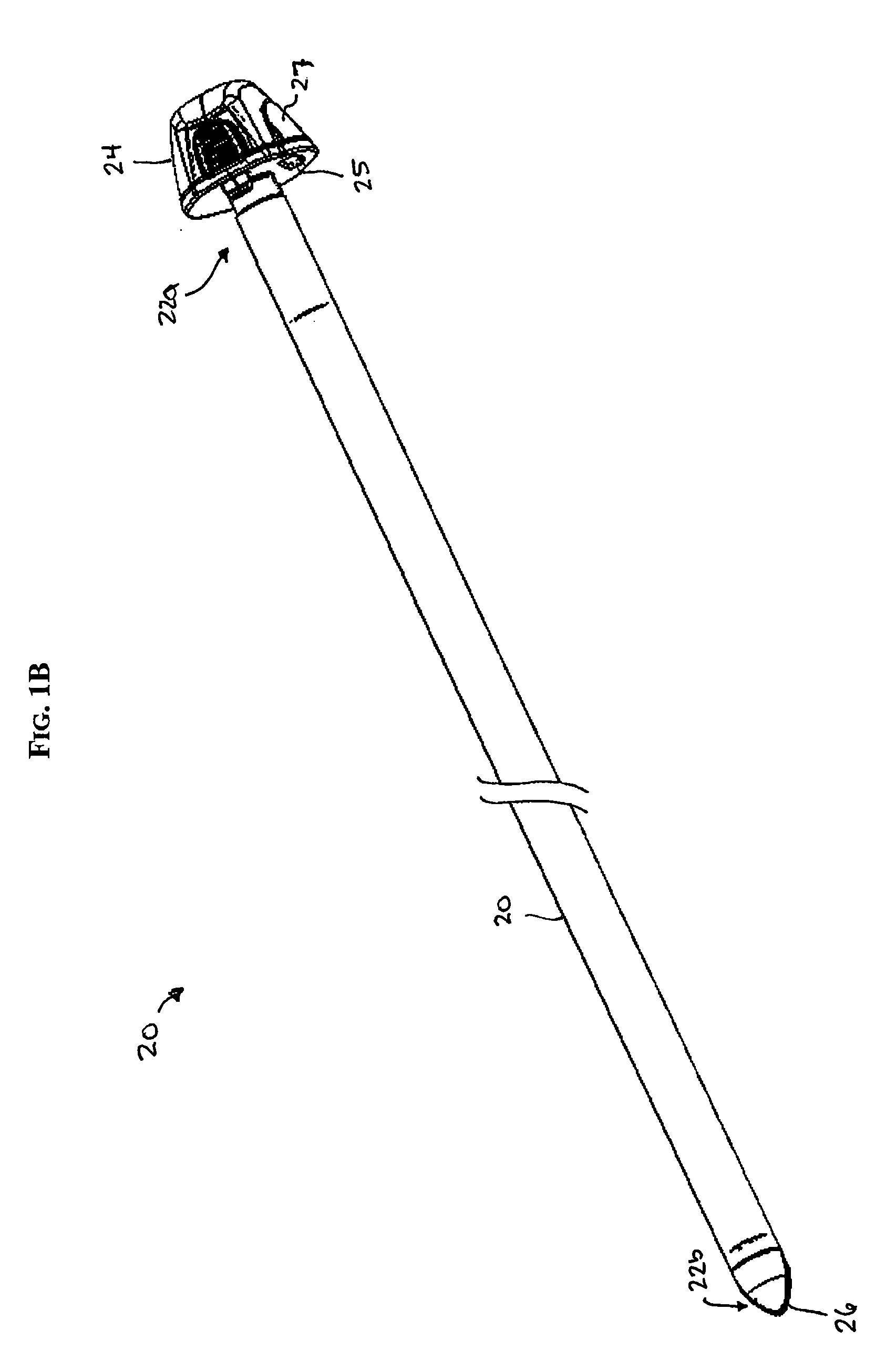
[0013] The trocar sleeve can also have a variety of configurations. In one embodiment, the trocar sleeve can include a tapered distal portion that tapers toward an outer surface of the end cap to form a substantially continuous outer surface with the end cap. The trocar sleeve can also optionally include at least one seal disposed therein which permits the passage of the end cap and an endoscope coupled to the end cap while limiting or preventing the passage of fluid or gas therethrough.
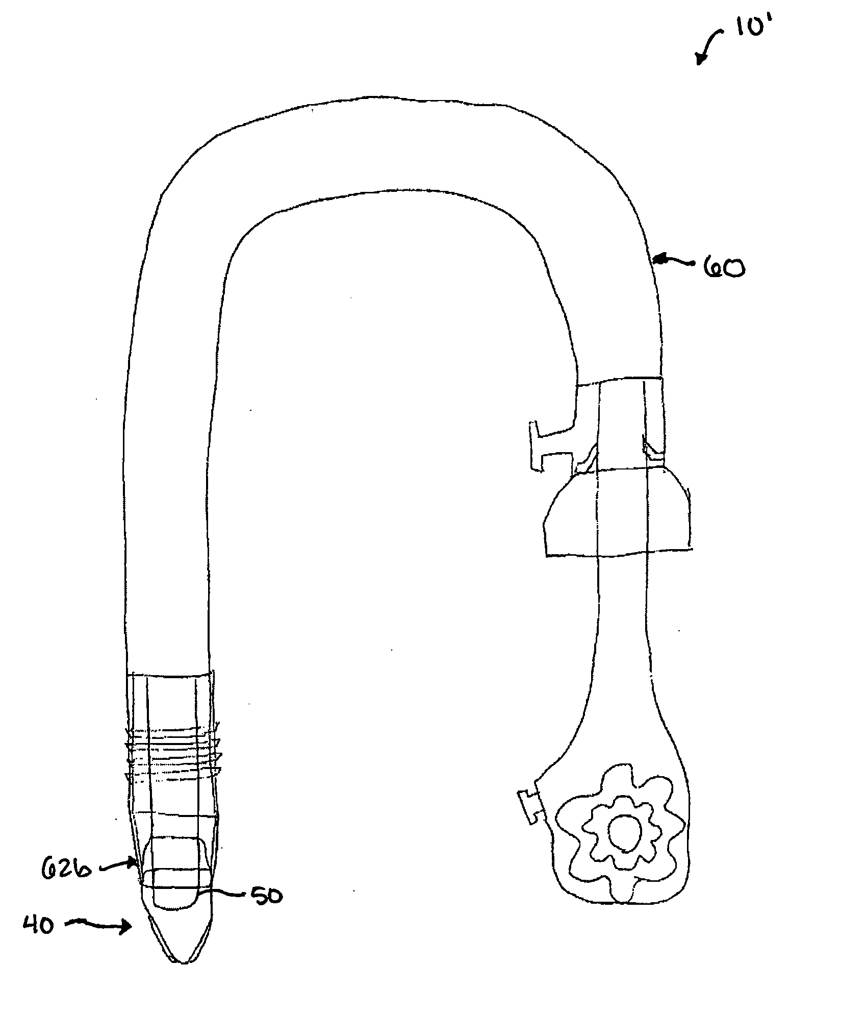
[0018] In one embodiment, advancing a portion of the sheath can include advancing at least one support rod between the proximal and distal ends of the sheath to advance a portion of the sheath into the body lumen. The body lumen can be, for example, an esophagus and the proximal end of the sheath can be disposed in an oral cavity at the opening of the esophagus. The portion of the sheath that is advanced translumenally can thus be advanced into a patient's stomach. The sheath will thus prevent direct contact between the endoscopic device and the esophagus. The method can also include positioning a distal end of the endoscopic device within a patient's stomach, inserting an endoscopic accessory through the endoscopic device, and using the endoscopic accessory to form a puncture hole in the stomach to access the patient's abdominal cavity. The endoscopic accessory can be, for example, a flexible trocar having a transparent distal tip shaped to penetrate and guide the flexible trocar sleeve through tissue to thereby insert the endoscopic device through tissue.
[0019] In another exemplary method for introducing an endoscopic device, a mid-portion of a sheath can be advanced through a body lumen and into a stomach cavity. The sheath can have proximal and distal ends that remain external to the body lumen while the mid-portion of the sheath is being advanced. An endoscopic device, with the distal end of the sheath coupled thereto, can then be advanced through the sheath to position a distal end of the endoscopic device within the stomach cavity. The sheath will prevent contact between the body cavity and the endoscopic device. In certain exemplary embodiments, advancing a mid-portion of the sheath can include advancing at least one support rod between the proximal and distal ends of the sheath to advance the mid-portion of the sheath into the body lumen.

Problems solved by technology

Obviously, the size and shape of instruments that may be passed through a bodily lumen in order to perform a medical procedure in the abdominal cavity are greatly restricted due to the anatomical properties of the lumen.
While current methods and devices used to penetrate tissue are effective, one drawback is that several exchanges and steps are required to form the opening.
In the event the endoscope is retracted through the opening, it can also be difficult to locate the opening and re-insert the endoscope.
Continued advancement and retraction of the endoscope can also be uncomfortable for the patient.
Another drawback of current methods is that a non-sterile passageway is created from the stomach into the abdominal cavity, as the devices carry bacteria from the body lumen into the abdominal cavity.

Method used


Image

  • Endoscopic Translumenal Surgical Systems
  • Endoscopic Translumenal Surgical Systems
  • Endoscopic Translumenal Surgical Systems

Examples


Embodiment Construction

[0038] Certain exemplary embodiments will now be described to provide an overall understanding of the principles of the structure, function, manufacture, and use of the devices and methods disclosed herein. One or more examples of these embodiments are illustrated in the accompanying drawings. Those of ordinary skill in the art will understand that the devices and methods specifically described herein and illustrated in the accompanying drawings are non-limiting exemplary embodiments and that the scope of the present invention is defined solely by the claims. The features illustrated or described in connection with one exemplary embodiment may be combined with the features of other embodiments. Such modifications and variations are intended to be included within the scope of the present invention.

[0039] The present invention generally provides methods and devices for performing translumenal (e.g., transoral and transanal) procedures. In general, the methods and devices utilize a tr...


PUM


Abstract

Methods and devices are provided for performing translumenal (e.g., transoral and transanal) procedures. In general, the methods and devices utilize a trocar assembly or trocar end cap that can facilitate insertion of an endoscope through tissue. In one embodiment, a flexible trocar assembly is provided and includes an obturator having an inner lumen formed therethrough for receiving an endoscope therein, and a distal end that is adapted to facilitate insertion of the endoscope through tissue and that is adapted to facilitate viewing therethrough. The trocar assembly can also include a trocar sleeve that is disposable over the obturator. In use, once the trocar assembly is inserted through tissue, the trocar sleeve can function as a placeholder, allowing the endoscope and obturator to be removed. The endoscope can then be removed from within the obturator and reinserted through the trocar sleeve for use in performing various other procedures. In other embodiments, rather than using a trocar that houses the endoscope, an end cap can be removably disposed over a distal end of the endoscope. The present invention also provides methods and devices for shielding an endoscope during insertion through a body lumen, and in particular for preventing contact between the endoscope (or trocar sleeve) and the body lumen, thus preventing bacteria from being carried into a body cavity.

Description

FIELD OF THE INVENTION [0001] The present invention relates to methods and devices for endoscopic translumenal surgery. BACKGROUND OF THE INVENTION [0002] Endoscopic surgery can be used to access the abdominal cavity via natural openings (mouth, anus, vagina, urethra) of the body and through the peritoneal lining of the abdominal cavity. Obviously, the size and shape of instruments that may be passed through a bodily lumen in order to perform a medical procedure in the abdominal cavity are greatly restricted due to the anatomical properties of the lumen. General surgeons, gastroenterologists, and other medical specialists, routinely use flexible endoscopes for intraluminal (within the lumen of the alimentary canal) examination and treatment of the upper gastrointestinal (GI) tract, via the mouth, and the lower GI tract, via the anus. In these procedures, the physician pushes the flexible endoscope into the lumen, periodically pausing to articulate the distal end of the endoscope usi...

Claims


Application Information

Patent Timeline
IPC IPC(8): A61B1/06
CPCA61B1/00142A61B17/3417A61B19/38A61B2017/2905A61B2017/00278A61B2017/00296A61B19/5212A61B90/361A61B90/40
Inventor BAKOS, GREGORY J.CROPPER, MICHAEL S.SMITH, RICHARD C.
Owner ETHICON ENDO SURGERY INC
Learn More