Signal acquisition and processing method and apparatus for magnetic resonance imaging

a magnetic resonance imaging and signal acquisition technology, applied in the field of medical imaging and image reconstruction, can solve the problems of adversely affecting system size and cost, high cost associated with such systems, and high cost associated with design and manufacture of such systems, and achieves the effects of low electrical noise level, maximum power reception, and high signal quality

Inactive Publication Date: 2007-11-22
TURSIOP TECH
View PDF2 Cites 5 Cited by
  • Summary
  • Abstract
  • Description
  • Claims
  • Application Information

AI Technical Summary

Benefits of technology

[0016] Conducting materials with interesting electrical properties, such as full or partial ballistic conductance, are used to make imaging coils in preferred embodiments of the invention. An example of such a material is a film drawn from carbon nanotubes. The relatively low electrical noise levels associated with such conductors permit the construction of RF receiving electronics circuitry with large Quality Factors, yielding maximum power reception, and thereby providing high signal quality and narrow bandwidth for highly selective imaging and optimal zone selection. The narrow bandwidth that can be attained thereby can be used for efficient imaging in the presence of an inhomogeneous static magnetic field, where slice selectivity for imaging is enhanced accordingly.
[0017] The invention teaches a method for making high-quality integrated imaging systems that are also relatively low in weight, in some cases so low in weight as to be portable.
[0018] In preferred embodiments, the imaging coils of the invention exploit ballistic conductance properties to optimize the B1 magnetic field within the imaging volume produced by the imaging coil per unit current flowing through the coil. In preferred embodiments of the invention, carbon nanotube conductors are made in thin film form with very small thickness at no cost in terms of increased resistance and are formed into geometrical shapes that create a relatively large B1 magnetic field within the imaging volume, while at the same time possessing only a small electrical resistance, thereby offering very high SNR values.

Problems solved by technology

A significant factor affecting further use of this versatile imaging technology is the high cost associated with such systems, both for purchase and maintenance.
The high costs associated with design and manufacture of such systems are due in large part to the necessity for a large and very homogeneous static magnetic field, as well as the need for gradient field producing apparatus for imaging with such systems; such large static fields are currently required in order to obtain high image quality and resolution.
However, the need for substantial homogeneity in the static magnetic field associated with a Magnetic Resonance Imaging system adversely impacts system size and cost.
Typically, current existing commercial methodologies are predicated on the use of a very homogeneous static magnetic field and cannot be used in the presence of inhomogeneous magnetic fields.
This large gradient magnitude leads to rapid dephasing of the spins after excitation by application of suitable Radio Frequency (RF) pulses.
As a practical matter, while these methods could be used to develop smaller imaging systems to image extremities or small peripheral portions of patient anatomy, they do not easily permit general internal imaging of patient anatomy.
Furthermore, the use of an imaging probe is invasive.
As such, these techniques do not permit general, non-invasive imaging within a wide field-of-view.

Method used


Image

  • Signal acquisition and processing method and apparatus for magnetic resonance imaging
  • Signal acquisition and processing method and apparatus for magnetic resonance imaging
  • Signal acquisition and processing method and apparatus for magnetic resonance imaging

Examples


Embodiment Construction

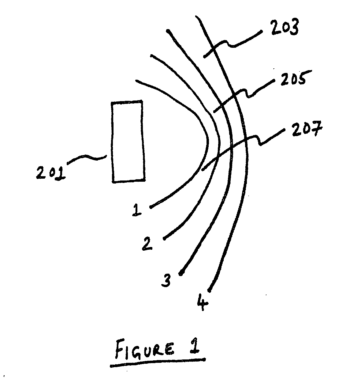
[0099] With reference to FIG. 1, in a preferred embodiment, a static magnetic field is generated by a single permanent magnet 201 (which could itself be a compound structure assembled from a variety of smaller magnets, with possibly varying magnetization directions) that generates a (typically inhomogeneous) magnetic field within a region of interest.

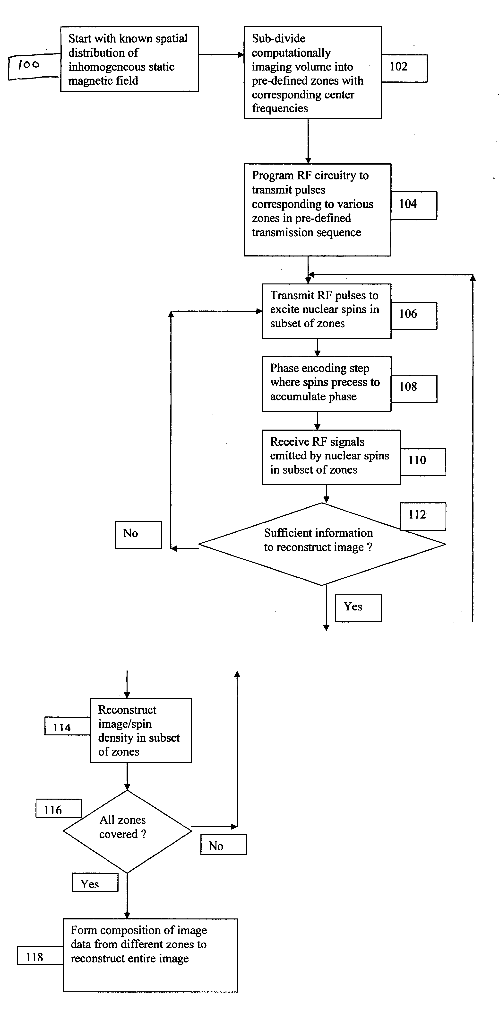
[0100] Standard Magnetic Resonance signal processing techniques are influenced by dephasing of the spins in the presence of inhomogeneities in the static magnetic field, leading to substantial signal decoherence or loss, whereupon image reconstruction becomes difficult or impossible. The teaching of the present invention provides a method of avoiding or greatly minimizing dephasing effects by taking into account the spatial variation of the static magnetic field, together with appropriate voxel shape selection and signal processing methodologies.

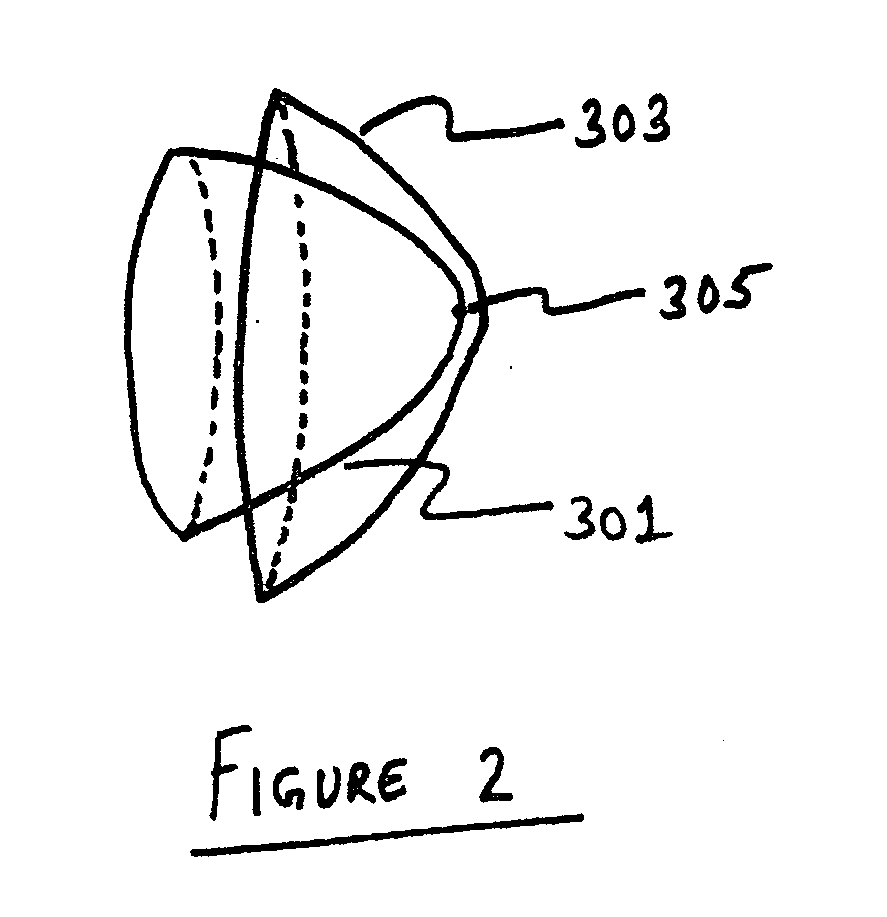
[0101] As shown in FIG. 1, the resulting magnetic field pattern can be divided into zones ...


PUM


Abstract

A method and apparatus are disclosed for Magnetic Resonance Imaging using specialized signal acquisition and processing techniques for image reconstruction with a generally inhomogeneous static magnetic field. New signal processing methods for image reconstruction and for minimizing dephasing effects are disclosed. Imaging systems with smaller static magnetic field strengths and smaller hardware demands than those with homogeneous static magnetic fields are provided, leading to significant reductions in system size and cost as compared to standard MRI systems. Such systems can also exploit imaging coils having high Signal-to-Noise-Ratio (SNR), including those made from Carbon nanotube conductors, leading to further imaging system efficiencies.

Description

CROSS-REFERENCES TO RELATED APPLICATIONS [0001] This application is entitled to the benefit of U.S. Provisional Patent Application No. 60 / 778,652, entitled “SIGNAL ACQUISITION AND PROCESSING METHOD AND APPARATUS FOR MAGNETIC RESONANCE IMAGING”, filed Mar. 4, 2006, herein incorporated by reference.FIELD OF THE INVENTION [0002] The present invention relates generally to medical imaging and image reconstruction, and specifically to Magnetic Resonance Imaging in the presence of static magnetic fields with an intrinsic field gradient. BACKGROUND OF THE INVENTION [0003] Magnetic Resonance (MR) Imaging technology is quite commonly used today in larger medical institutions worldwide, and has led to huge benefits in the practice of medicine. A significant factor affecting further use of this versatile imaging technology is the high cost associated with such systems, both for purchase and maintenance. [0004] The high costs associated with design and manufacture of such systems are due in larg...

Claims


Application Information

Patent Timeline
Patent Type & Authority Applications(United States)
IPC IPC(8): G01V3/00
CPCG01R33/4833G01R33/3808
Inventor VISWANATHAN, RAJU
Owner TURSIOP TECH
Why Eureka
Try Eureka