
System and Micro-Catheter Devices for Medical Imaging of the Breast

a micro-catheter device and breast technology, applied in the field of breast medical imaging system and micro-catheter devices, can solve the problems of no teaching of inserting a ductascope into the nipple, no previous work discussing the unique abilities of holey fibers, and the failure of ductoscopy

Inactive Publication Date: 2007-11-22
SCHELLENBERG JAMES
View PDF9 Cites 10 Cited by
  • Summary
  • Abstract
  • Description
  • Claims
  • Application Information

AI Technical Summary

Problems solved by technology

However, there is no teaching of inserting a ductascope into the nipple while the cup is in place.
As well, none of the previous work discusses the unique abilities of holey fiber as applied to micro-catheter design.
This paper also noted that nipple manipulation was required to access some of the ducts, and that the majority of the ducts were not so convoluted that ductoscopy would not work.

Method used


Image

  • System and Micro-Catheter Devices for Medical Imaging of the Breast
  • System and Micro-Catheter Devices for Medical Imaging of the Breast
  • System and Micro-Catheter Devices for Medical Imaging of the Breast

Examples


Embodiment Construction

[0042] Unless defined otherwise, all technical and scientific terms used herein have the same meaning as commonly understood by one of ordinary skill in the art to which the invention belongs. Although any methods and materials similar or equivalent to those described herein can be used in the practice or testing of the present invention, the preferred methods and materials are now described. All publications mentioned hereunder are incorporated herein by reference. As used herein, the terms “channel” and “lumen” are in some contexts used interchangeably as will be readily apparent to one of skill in the art.

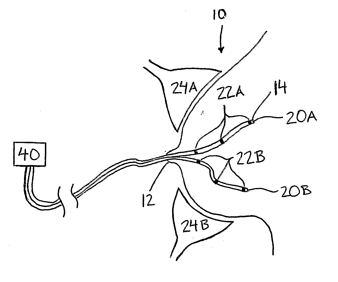
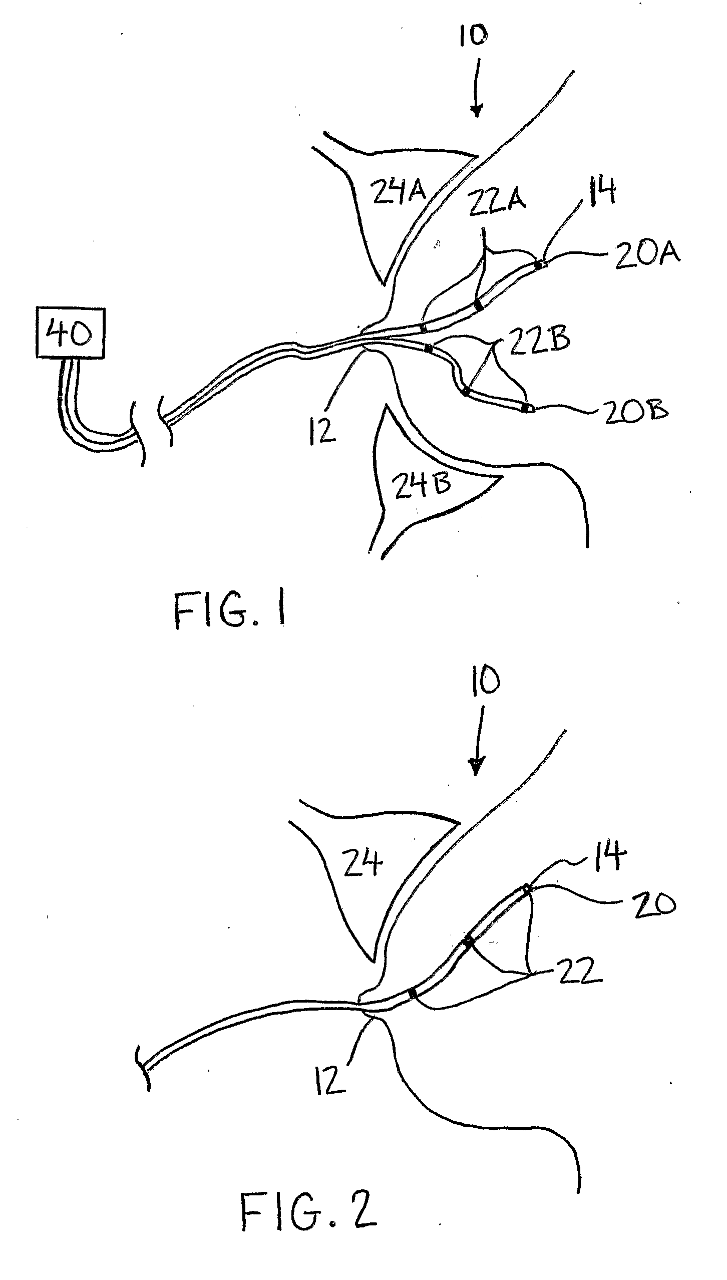
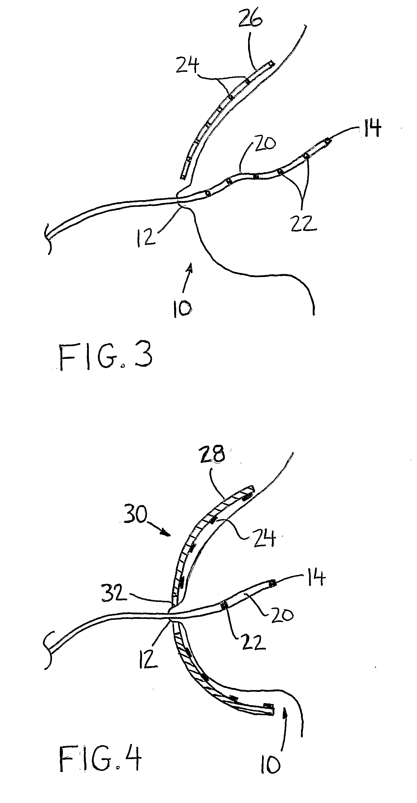
[0043] Described herein is a method of scanning breast tissue wherein an imaging element is inserted within a breast via a carrier inserted into a breast duct within the breast for imaging the breast tissue located between the in-duct imaging element and an imaging element located outside of the breast. As will be appreciated by one of skill in the art, inserting an imaging ele...


PUM


Abstract

A method of scanning breast tissue involves inserting an imaging element into a breast via a carrier inserted into a breast duct. The imaging element may be for example a receiver, a transmitter or a mirror. A signal is transmitted between one imaging element within the breast duct and a second imaging element outside the breast.

Description

PRIOR APPLICATION INFORMATION [0001] This application claims the benefit of U.S. Provisional Patent Application 60 / 577,167, filed Jun. 7, 2004.BACKGROUND OF THE INVENTION [0002] Holey Fiber, also called Crystal Fiber, was first developed at the University of Bath in 1995 based on the work of Philip Russell. Unlike normal optical fiber which is solid throughout, holey fiber is fabricated with one or more continuous open channels running from one end to the other. These can be very small diameter channels, with diameters of 3 micrometers and less being possible today. Holey fiber may be made from glass or plastic. These channels have previously been filled with gas in an application to develop a high-power laser. Recently, holey fiber has been investigated for use in medical applications within the endoscope subject area, in which viewing of the interior of the body is the purpose (http: / / www.newscientist.com / article.ns?id=dn4589). This work was done by Martijn van Eijkelenborg, Austr...

Claims


Application Information

Patent Timeline
Patent Type & Authority Applications(United States)
IPC IPC(8): A61B1/00A61B5/00A61B5/06
CPCA61B5/0062A61B5/0084A61B5/0086A61B5/68A61B5/0507A61B5/06A61B5/4312A61B5/0091A61B5/065
Inventor SCHELLENBERG, JAMES
Owner SCHELLENBERG JAMES
Learn More