Cell Growth Inhibiting Film, Medical Instrument and Digestive System Stent

a technology of cell growth and inhibiting film, which is applied in the direction of prosthesis, packaging foodstuffs, and packaged goods, etc., can solve the problems of imposing a large burden on the patient and serious symptoms such as pancreatitis, and achieve the effect of preventing side effects due to the physiologically active substan

Inactive Publication Date: 2007-11-29
ZEON MEDICAL
View PDF4 Cites 11 Cited by
  • Summary
  • Abstract
  • Description
  • Claims
  • Application Information

AI Technical Summary

Benefits of technology

[0010] The inventors of the present invention prepared a film having a porous honeycomb structure by casting an organic solvent solution of a resin such as 1,2-polybutadiene onto a substrate using a method similar to the method disclosed in JP-A-2002-335949 and JP-A-2003-149096. The inventors placed the resulting film in a culture medium and cultured malignant gallbladder carcinoma cells on the film. Surprisingly, the inventors have found that the growth of the cancer cells was remarkably inhibited in contrast to the example using cardiac muscle cells disclosed in JP-A-2002-335949. The inventors also have found that a medical instrument which does not impose a burden on the patient due to side effects caused by a physiologically active substance and can inhibit the progress of cancer can be obtained by covering a medical instrument substrate with the above film.
[0028] According to the cell growth inhibiting film, the cell growth inhibiting method, and the medical instrument of the present invention, since the cell growth inhibitory effects can be obtained without using a physiologically active substance, side effects due to the physiologically active substance can be prevented.

Problems solved by technology

However, this medical instrument has a problem in which the physiologically active substance causes significant side effects on the human body to impose a large burden on the patient.
The covered stent is useful for preventing stricture of a tubular cavity in the body due to the growth of cancer cells or the like, since the resin film does not allow the cancer cells to pass through.
However, since the film used for the covered stent cannot allow a digestive fluid such as a pancreatic juice to pass through, the flow of the digestive fluid is hindered due to the covered stent, whereby a serious symptom such as pancreatitis may occur.

Method used


Image

  • Cell Growth Inhibiting Film, Medical Instrument and Digestive System Stent
  • Cell Growth Inhibiting Film, Medical Instrument and Digestive System Stent
  • Cell Growth Inhibiting Film, Medical Instrument and Digestive System Stent

Examples


example 1

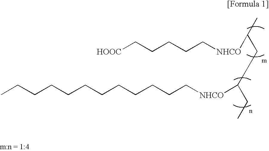
[0114] 6 ml of a solution (resin concentration: 0.27 wt %) prepared by dissolving poly(ε-caprolactone) (manufactured by Wako Pure Chemicals Co., Ltd., viscosity average molecular weight: 70,000) and a Cap resin (weight average molecular weight: 62,000, number average molecular weight: 21,000) in chloroform at a weight ratio of 10:1 was uniformly spread on a glass petri dish with a diameter of 10 cm.

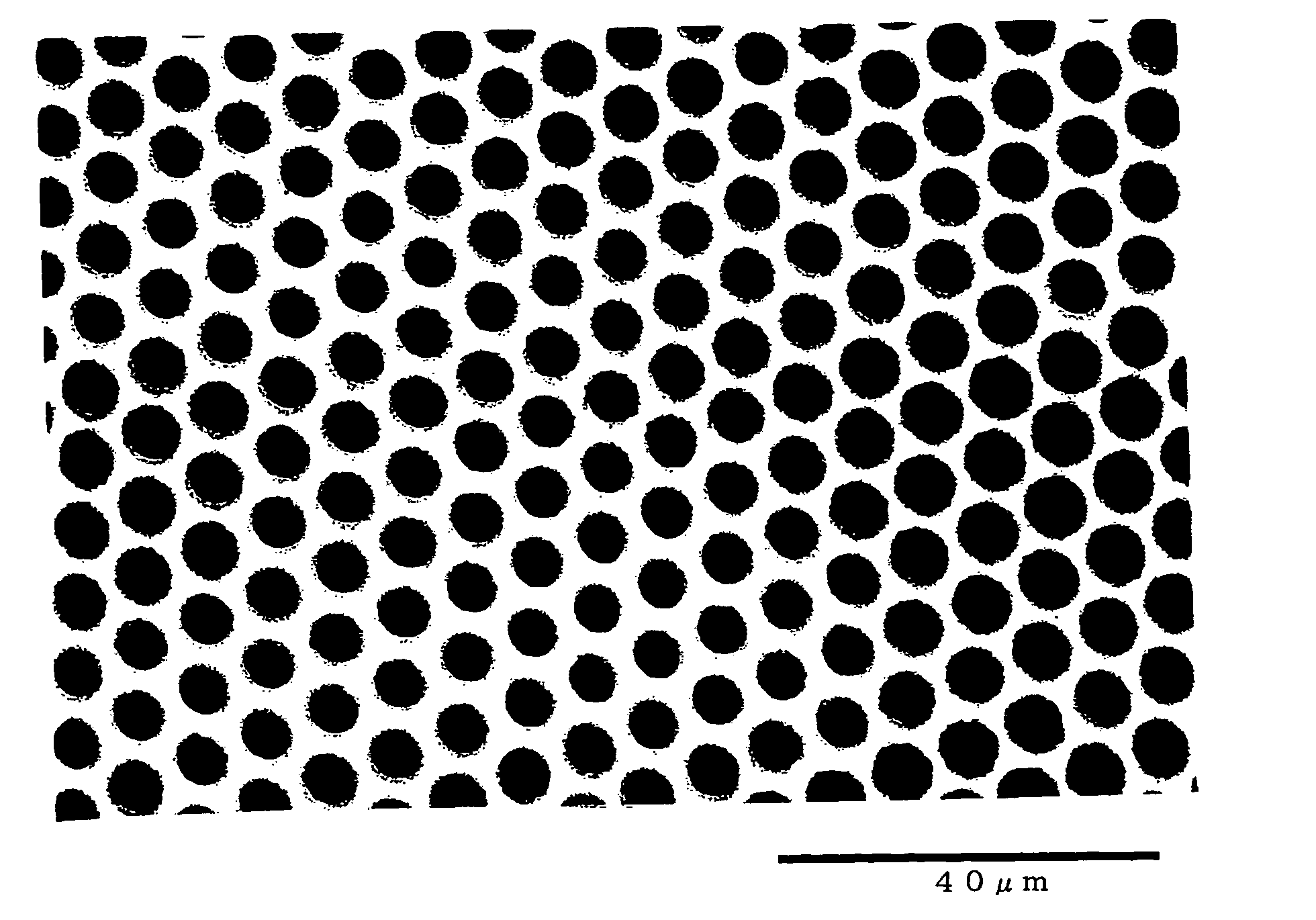
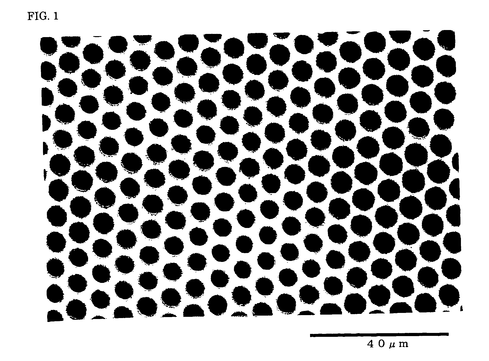
[0115] Then, high-humidity air with a relative humidity of 70% was sprayed onto the liquid surface on the glass petri dish for one minute at a flow rate of 2 l / min in an atmosphere at a temperature of 23.0° C. and a relative humidity of 40% to obtain a film A with a thickness of 1 to 2 μm. The film A was observed using an optical microscope (“BH2” manufactured by Olympus Corporation) at a magnification of 100. It was confirmed that a porous honeycomb structure was formed in the film A. The pores of the porous structure had an average pore size of 3.5 μm and a coefficient of variation in ...

examples 2 and 3

[0116] In Examples 2 and 3, films B and C having a porous honeycomb structure were obtained in the same manner as in Example 1 except for changing the temperature of the atmosphere to 24.0° C. and 25.0° C., respectively.

[0117] Table 1 shows the thicknesses of the films B and C and the average pore size and the coefficient of variation in pore size of the pores of the porous structure.

examples 4 to 6

[0118] Films D, E, and F were respectively obtained in the same manner as in Examples 1, 2, and 3 except for using 1,2-polybutadiene (“RB820” manufactured by JSR Corporation) as the resin instead of poly(F-caprolactone).

[0119] As a result of observation using an optical microscope, it was confirmed that a porous honeycomb structure was formed in the films D to F. Table 1 shows the thicknesses of the films D to F and the average pore size and the coefficient of variation in pore size of the pores of the porous structure.


PUM

PropertyMeasurementUnit
pore sizeaaaaaaaaaa
pore sizeaaaaaaaaaa
pore sizeaaaaaaaaaa

Abstract

A cell growth inhibiting film including a resin and having a porous structure formed at least on its surface, a cell growth inhibiting method including causing the surface of a film including a resin and having a porous structure formed at least on its surface to contact cells to inhibit growth of the cells in the contact area, a medical instrument including a medical instrument substrate and a film including a resin and having a porous structure formed at least on its surface, in which the surface of the medical instrument substrate is entirely or partially covered with the film, and a digestive system stent including a stent substrate and a film including a resin and having a porous structure formed by through-holes with an average pore size of 0.1 to 20 μm and a coefficient of variation in pore size of 30% or less, in which the stent substrate is covered with the film. According to the present invention, a material exhibiting cell growth inhibitory effects without using a physiologically active substance and suitable for forming a medical instrument, and a digestive system stent which secures a digestive system tubular cavity in the body and allows a digestive fluid and digestive enzymes contained therein to pass through, but blocks cancer cells, can be provided.

Description

TECHNICAL FIELD [0001] The present invention relates to a cell growth inhibiting film, a cell growth inhibiting method using the cell growth inhibiting film, a medical instrument, and a digestive system stent which is placed in a digestive system tubular cavity in the body such as the bile duct, esophagus, duodenum, or large intestine. BACKGROUND ART [0002] In regard to the interaction between cells and a material, it is known that the cells are affected not only by the chemical properties of the surface of the material, but also by the minute shape of the surface of the material. For example, JP-A-2002-335949 discloses a film or a stretched film having a honeycomb structure which is obtained by casting a hydrophobic organic solvent solution of a biodegradable and amphiphilic polymer or a polymer mixture of a biodegradable polymer and an amphiphilic polymer onto a substrate, causing the organic solvent to be evaporated and condensed on the surface of the cast organic solvent solutio...

Claims


Application Information

Patent Timeline
Patent Type & Authority Applications(United States)
IPC IPC(8): A61L31/00A61L27/00A61L31/14
CPCA61L31/146
Inventor TANAKA, MASARUSHIMOMURA, MASATSUGUTOYOKAWA, YOSHIHIDE
Owner ZEON MEDICAL
Why Eureka
Try Eureka