Method and processor for generating a medical image

Active Publication Date: 2008-02-14
SIEMENS HEALTHCARE GMBH
View PDF15 Cites 23 Cited by
  • Summary
  • Abstract
  • Description
  • Claims
  • Application Information

AI Technical Summary

Benefits of technology

[0026] The magnification factor and/or the image section to be depicted are thus also used in further image processing steps. For example, a segmentation algorithm that is based on the magnification factor and/or the image section

Problems solved by technology

If a user must sequentially consider many medical images (as is the case, for example, for digital slice image data sets that have been acquired with by computed tomography o

Method used


Image

  • Method and processor for generating a medical image
  • Method and processor for generating a medical image
  • Method and processor for generating a medical image

Examples


Embodiment Construction

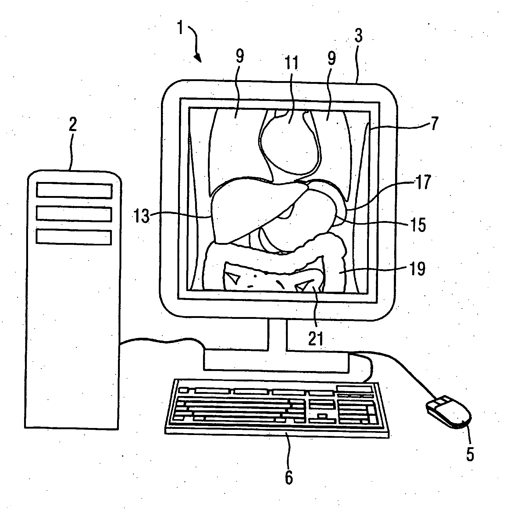
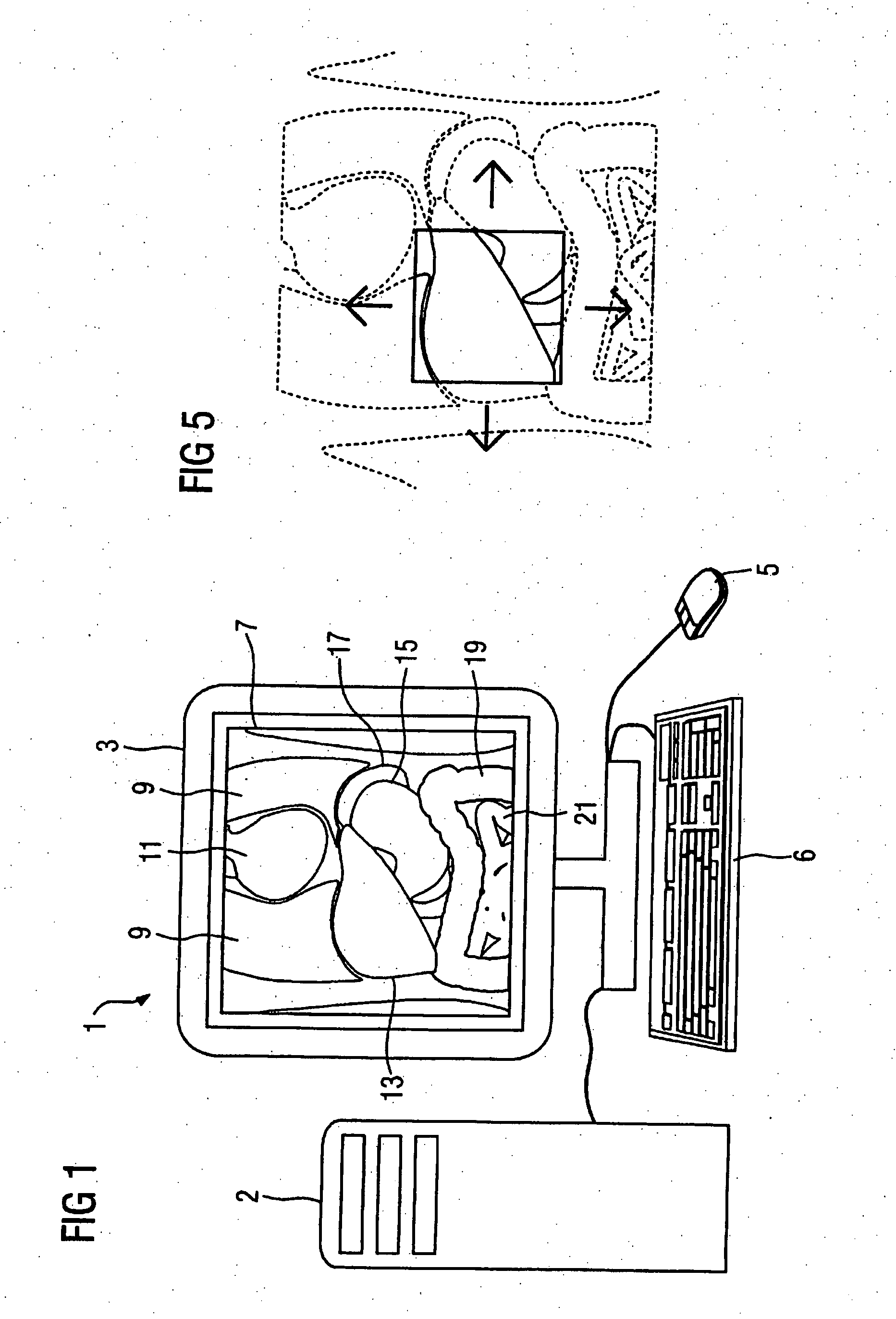
[0032]FIG. 1 shows a presentation unit 1 for medical images. Such a presentation unit 1 typically includes a monitor 3 with which medical images are shown to a user as well as input means (for example a keyboard 6 or a mouse 5) with which a user can vary the presentation of a medical image and adjust it according to his wishes. The presentation unit 1 is connected with a computer 2 that includes means for administration of or for connecting to a databank so that acquired data sets as well as information linked therewith (such as, for example, examination indications, patient data, acquisition modalities) can be loaded or stored and means for processing of the data so that embodiments of the inventive method as they are subsequently described can be executed.

[0033] As an example and for explaining the inventive method, a frontal section 7 through an epigastrium of a patient to be examined is shown to a user with the aid of the presentation unit 1. Such a frontal section 7 can be acq...


PUM


Abstract

In a method for generation of a medical image, starting from an acquired data set of an organ to be depicted, a magnification factor and/or an image section to be depicted are automatically determined dependent on the organ to be depicted. Based on this a medical image is generated from the data set. A data processing unit for medical images is fashioned for implementation of such a method and computer software implements such a method when it is executed on a data processing unit.

Description

BACKGROUND OF THE INVENTION [0001] 1. Field of the Invention [0002] The invention concerns a method for generation of a medical image, as well as a data processing unit and computer software for this purpose. [0003] 2. Description of the Prior Art [0004] In the examination of medical images a user (for example, a physician) must often review and assess many images in the shortest possible time. A user thus will typically predominately direct his or her attention to an organ for which assessment is of central importance for evaluation of a medical question. Since the relevant medical information is often found in small regions of the medical image and since the medical images today for the most part exist digitally, a user will look at corresponding image sections in an enlarged view for better assessment of this information. If a user must sequentially consider many medical images (as is the case, for example, for digital slice image data sets that have been acquired with by compute...

Claims


Application Information

Patent Timeline
IPC IPC(8): A61B5/00
CPCG06T7/0012G06T2207/30004G06T2207/10072
Inventor ASSMANN, STEFANEKINCI, OKAN
Owner SIEMENS HEALTHCARE GMBH
Why Eureka
Try Eureka