Ultrasonic Diagnostic Apparatus

a diagnostic apparatus and ultrasonic technology, applied in the field of ultrasonic diagnostic equipment, can solve the problems of reducing the diagnostic efficiency, the degree of hardness of the displayed tissues compared to the surrounding tissues cannot be quantified, etc., and achieve the effect of improving the diagnostic efficiency

Inactive Publication Date: 2008-02-28
HITACHI MEDICAL CORP
View PDF23 Cites 40 Cited by
  • Summary
  • Abstract
  • Description
  • Claims
  • Application Information

AI Technical Summary

Benefits of technology

[0014] The objective of the present invention is to provide an ultrasonic diagnostic apparatus that makes it possible for a doctor to grasp hardness of the affected area by being able to observe the strain image or elastic module image more quantitatively compared to the prior art, and improves efficiency of diagnosis thereof.

Problems solved by technology

However, degree of hardness of the displayed tissues comparing to the surrounding tissues could not be acknowledged quantitatively.
Also, even though a doctor can specify the hue of the target region in the strain image to which the hue information is assigned since the hue of color bars are displayed with gradation, they have to spend much time for specifying where the hue falls in the color bar which lowers the efficiency of diagnosis.

Method used


Image

  • Ultrasonic Diagnostic Apparatus
  • Ultrasonic Diagnostic Apparatus
  • Ultrasonic Diagnostic Apparatus

Examples


Embodiment Construction

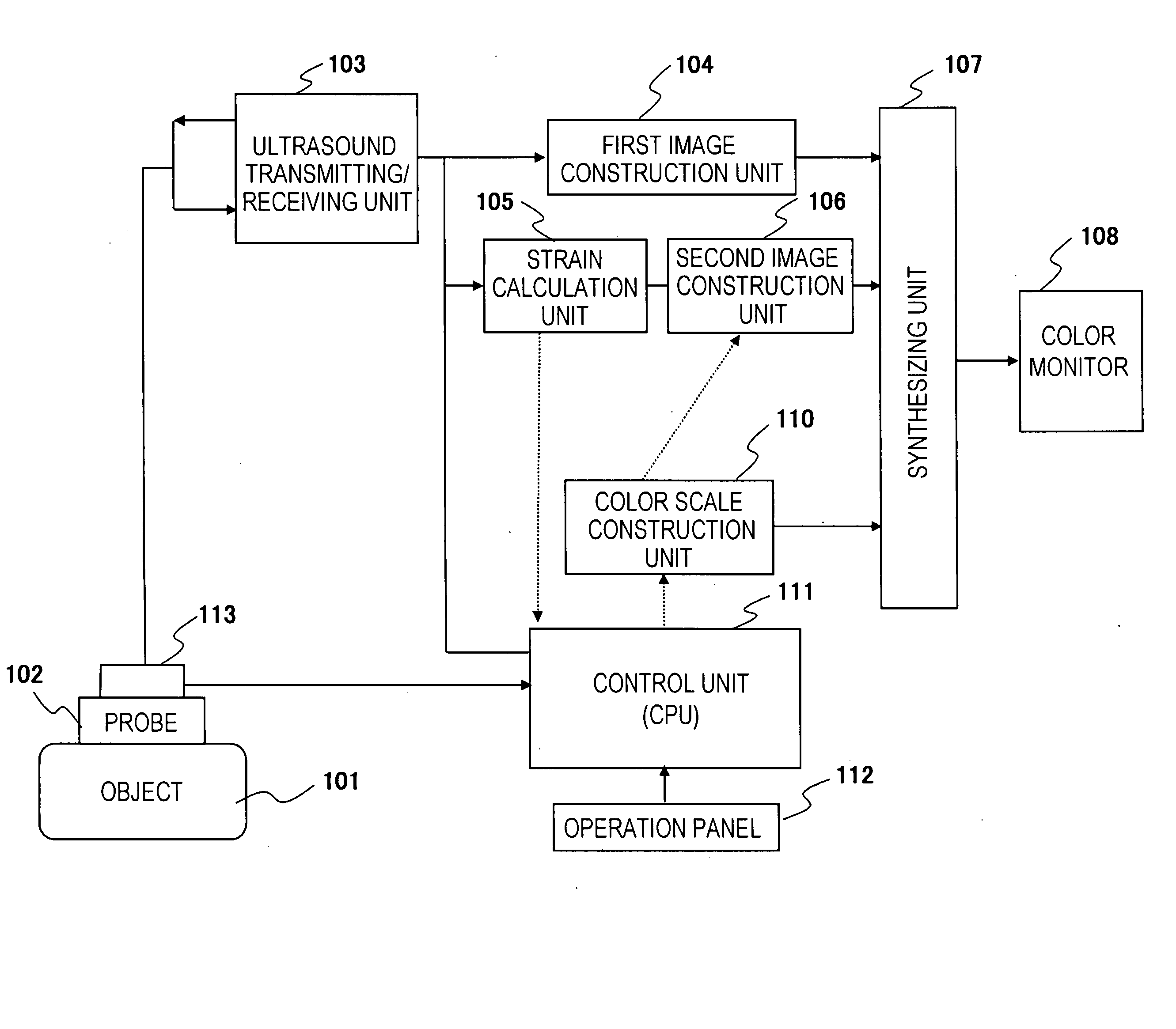
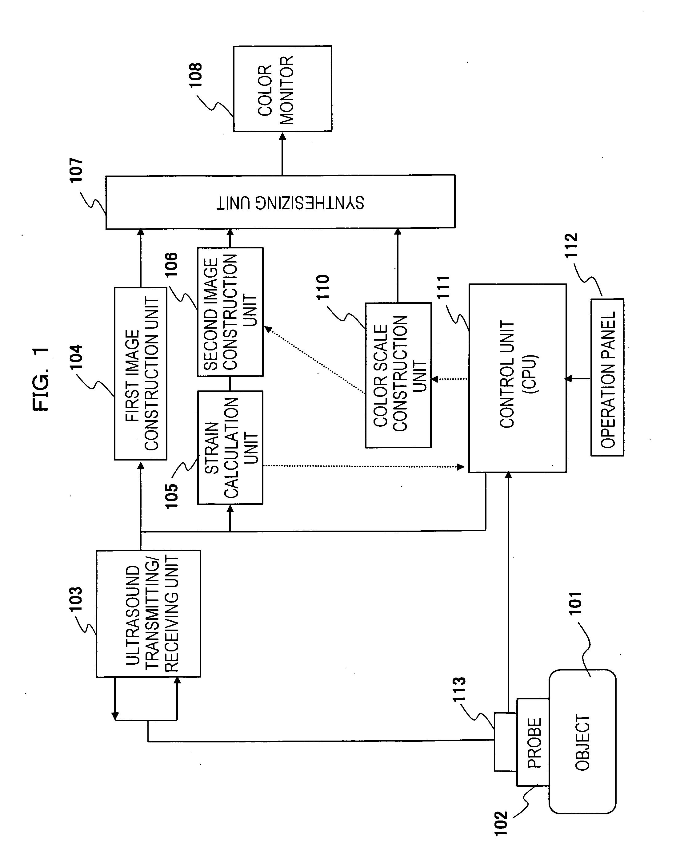
[0025] Hereinafter, embodiments of the present invention will be described referring to the diagrams. As seen in FIG. 1, an ultrasonic diagnostic apparatus to which the present invention is applied comprises:

[0026] ultrasound probe 102 for applying to object 101, and transmitting ultrasonic beams to object 101 as well as receiving ultrasonic waves reflected in the body of object 101;

[0027] a transmitting circuit for providing transmitting signals that transmit ultrasonic waves to object 101 with a predetermined time interval;

[0028] a receiving circuit for receiving echoes reflected in the body of object 101, converting them into electronic signals (echo signals) and outputting them;

[0029] ultrasound transmitting / receiving unit 103 provided with a phasing addition circuit for forming ultrasonic beam signals (RF signal data) by executing phasing addition process on the echo signals being outputted from the receiving circuit, and outputting them;

[0030] first image construction uni...


PUM


Abstract

An ultrasonic diagnostic apparatus (ultrasonic elastograph) for measuring strain or elastic modulus of a tissue or an organ of a living body, creating a strain image or an elastic modulus image by assigning a hue to the measurement data corresponding to the measured value, combining these images with a black and white tomogram, displaying the synthetic image on a color monitor, and displaying the hue assigned to the measurement data with a color bar. Information on comparison between a measured value corresponding to the hue of the color bar displayed on the color monitor and the reference value of the measured value is displayed at a position adjacent to the color bar on the screen of the color monitor.

Description

TECHNICAL FIELD [0001] The present invention relates to an ultrasonic diagnostic apparatus, in particular, to the ultrasonic diagnostic apparatus for creating a strain image of an organ in a living body that contributes to medical diagnosis. BACKGROUND ART [0002] The ultrasonic diagnostic apparatuses transmit ultrasonic waves to the inside of the body of an object to be examined, detect echoes reflected from the body tissues, create and display images of those reflected signals. Images displayed in these apparatuses are tomograms presenting tissue characterization of the body of the object which is measured approximately in real time by applying an ultrasound probe on the surface of the object, or images presenting blood flow or movement of organs being measured with application of Doppler effect. [0003] Upon displaying images of blood flow or movement of organs using an ultrasonic diagnostic apparatus, blood flow or movement of organs is color displayed on the monitor with black an...

Claims


Application Information

Patent Timeline
Patent Type & Authority Applications(United States)
IPC IPC(8): A61B8/00A61B8/08A61B8/13G01S7/52
CPCA61B8/08A61B8/13G01S7/52071G01S7/52042A61B8/463
Inventor WAKI, KOJIMURAYAMA, NAOYUKI
Owner HITACHI MEDICAL CORP
Why Eureka
Try Eureka