
Methods and apparatuses for medical imaging

a technology of medical imaging and apparatus, applied in the field of medical imaging, can solve the problems of labor-intensive and subjective providing suitable background samples, difficulty for human observers to distinguish the boundary between blood and the vessel wall, and computational cos

Inactive Publication Date: 2008-02-28
UNIV HOUSTON SYST
View PDF62 Cites 149 Cited by
  • Summary
  • Abstract
  • Description
  • Claims
  • Application Information

AI Technical Summary

Benefits of technology

The present invention provides a method for medical imaging that can produce anatomical, physiological, and morphological data about a vessel and its structures, including plaque development and inflammation. The method can be used on animals and humans. The invention also provides catheters with a contrast agent delivery system and a Doppler sensor for simultaneous IVUS imaging and Doppler blood flow imaging of regions of interest. The invention also provides methods for detecting and quantifying regions of contrast perfusion into a vessel wall, visualizing micro-vascularized plaque and adventitia segments of a vessel, and visualizing vasa vasorum. Overall, the invention enhances the ability to diagnose and treat vascular disease.

Problems solved by technology

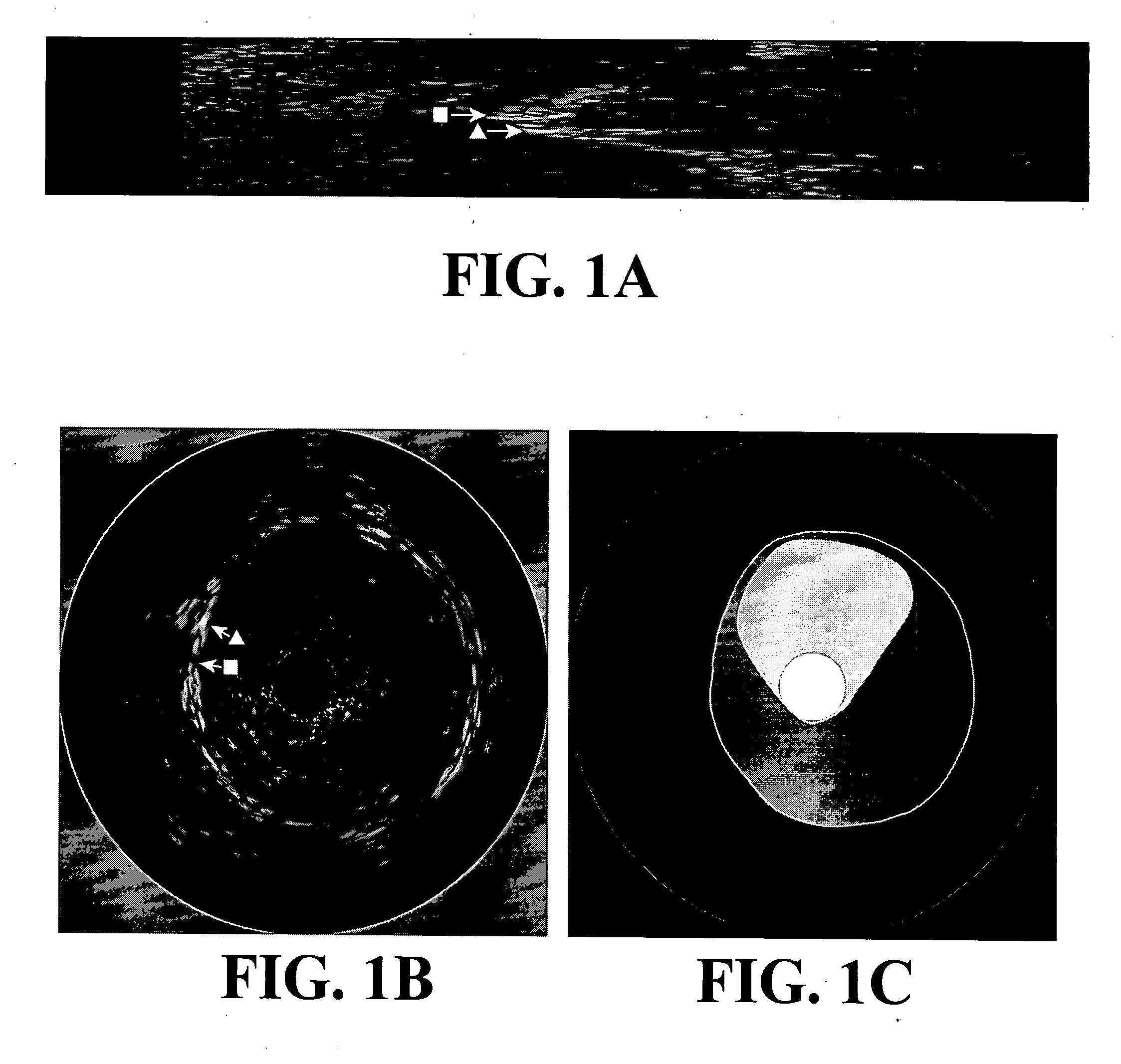
While the benefits obtained from improved image resolution often outweigh the other issues, the problem of blood backscatter is of particular concern as it makes it difficult for a human observer to distinguish the boundary between the blood and the vessel wall.
This contributes to the known problems associated with the reproducibility of vessel morphology studies [1].
Providing suitable background samples may be labor-intensive and subjective.
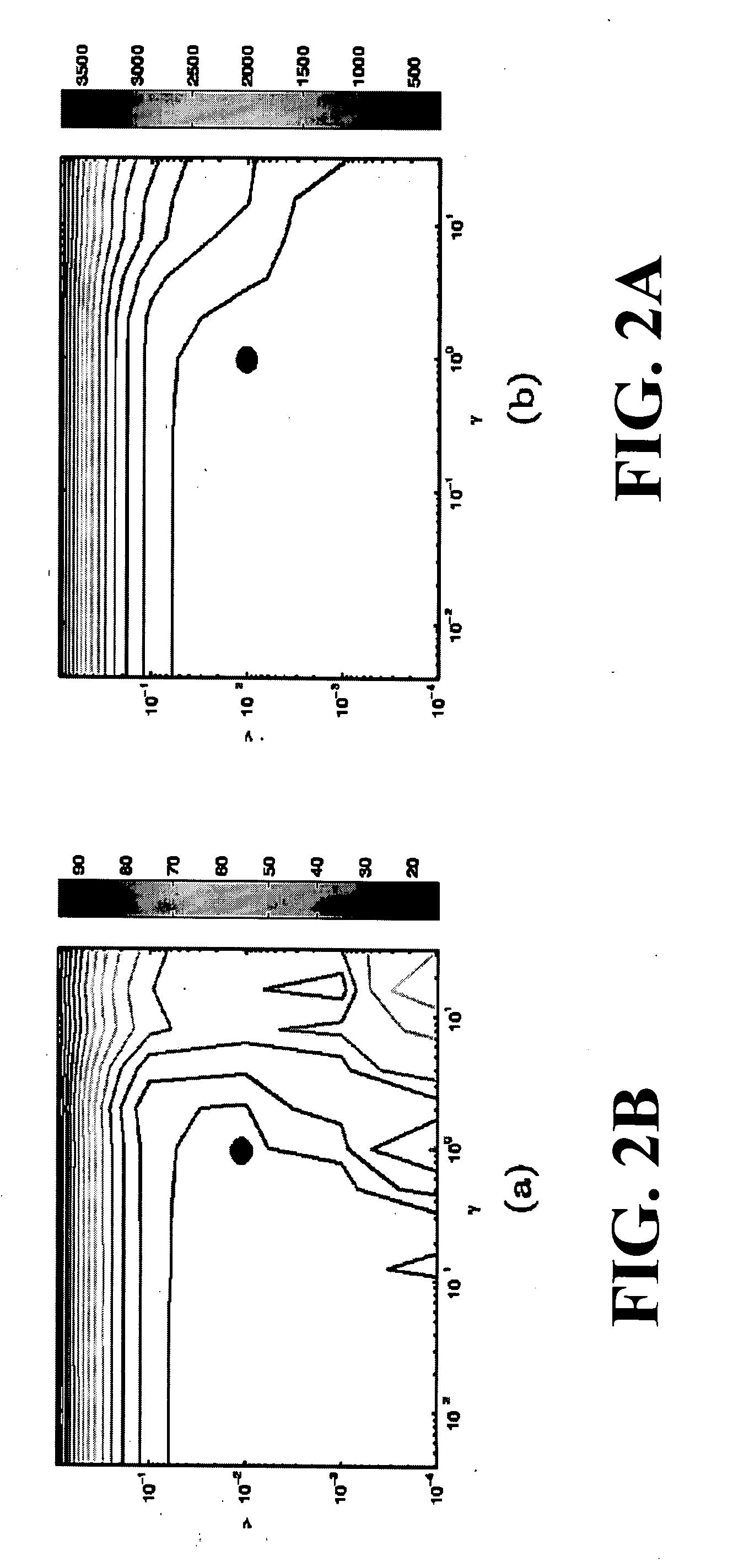
A high number of support vectors relative to the number of training examples is not only indicative of overfitting, but is computationally expensive when it comes to later recognizing a sample of unknown class.
For the purposes of this study we ignore the adventitia and surrounding tissues: they not only frequently contain free blood themselves, but are often very difficult for a human observer to reproducibly interpret.
More elaborate feature selection schemes such as genetic algorithms [7] could be used, but as one of our goals here is to determine which feature(s) best characterize the blood, we will not investigate this issue.
(Note that a temporal window of tθ=13 may be excessively long for an IVUS system whose frame rate is below 30 frames / s.)

Method used


Image

  • Methods and apparatuses for medical imaging
  • Methods and apparatuses for medical imaging
  • Methods and apparatuses for medical imaging

Examples


Embodiment Construction

[0049] The inventors have developed a number of systems, apparatuses and method for improving data derived from intravascular ultrasound. These systems, apparatuses and methods include: (A) new catheter designs including contrast agent introduction subsystems and / or Doppler subsystems; (B) methods for acquiring and analyzing Doppler data from intravascular ultrasound (IVUS) catheters; (C) method for RF-based detection of blood and / or contrast agents such as micro-bubbles; (D) methods for frame-grating image data analysis, (E) methods for difference imaging for contrast detection; (F) methods for quantification and visualization; and (G) methods for performing IVUS imaging.

A. Catheter Design

[0050] The present invention also relates to a contrast enhanced IVUS (CEIVUS) catheter and / or Doppler enhanced IVUS catheter. The catheter includes a nozzle system having exit holes disposed around its periphery, where the holes are adapted to direct jets of a contrast agent near, immediately p...


PUM


Abstract

A set of intravascular ultrasound (IVUS) related systems, apparatuses and methods are disclosed. New catheter designs including contrast agent introduction subsystems and / or Doppler subsystems are disclosed. Methods for acquiring and analyzing Doppler data from intravascular ultrasound (IVUS) catheters are disclosed. RF-based detection of blood and / or contrast agents such as micro-bubbles are disclosed. Methods for frame-grating image data analysis permitting frame registration before, during and after a contrasting effect is imposed on a system being imaged are disclosed. Methods for difference imaging for contrast detection are disclosed. Methods for quantification and visualization of IVUS data are disclosed. And methods for IVUS imaging are disclosed.

Description

RELATED APPLICATIONS [0001] This application is a continuation-in-part of U.S. patent application Ser. No. 10 / 586,020, filed Jul. 14, 2006, which is a 371 nationalized application of PCT Patent Application Ser. No. PCT / US05 / 01436, filed Jan. 14, 2005, which claims priority U.S. patent Provisional Patent Application Ser. No. 60 / 536,807, filed Jan. 16, 2004.GOVERNMENTAL INTEREST [0002] Governmental entities may have certain rights in and to the contents of this application due to funded from NSF Grant IIS-0431144 and a NSF Graduate Research Fellowship (SMO).BACKGROUND OF THE INVENTION [0003] 1. Field of the Invention [0004] The present invention relates to methods and systems for detecting and localizing vasa vasorum or other microvessels or micro-vascularizations associated with arteries, veins, tissues, organs and cancers in animals including humans. [0005] More particularly, the present invention relates to methods and systems including the steps of acquiring contrast-enhanced data...

Claims


Application Information

Patent Timeline
Patent Type & Authority Applications(United States)
IPC IPC(8): A61B8/06
CPCA61B8/06A61B8/0833A61B8/12A61B8/469A61B8/481G06K9/522A61B8/543G01S7/52066G01S7/52071G01S15/8979A61B8/488G06V10/431
Inventor KAKADARIS, IOANNIS A.NAGHAVI, MORTEZAO'MALLEY, SEAN M.
Owner UNIV HOUSTON SYST
Learn More