Computer aided diagnosis using video from endoscopes

a technology of endoscopy and computer aided diagnosis, applied in the field of medical imaging, can solve the problems of inability to distinguish between polyps or lesions and residual stool or other material in the colon, incurring greater risks of bleeding, infection and other adverse side effects, and scans that do not provide information about the color or texture of the interior surface of the colon, etc., to achieve the effect of enhancing the image fram

Inactive Publication Date: 2008-03-06
CADES SCHUTTE A LIMITED LIABILITY LAW PARTNERSHIP
View PDF16 Cites 159 Cited by
  • Summary
  • Abstract
  • Description
  • Claims
  • Application Information

AI Technical Summary

Problems solved by technology

Further, the colonoscopist must locate the actual polyp or lesion (which was shown in the scan) in the patient's colon at a remote time after the scan, which can be very difficult.
Also, scans do not provide information about the color or texture of the interior surface of the colon, which would provide diagnostic information about vessel structure and pit patterns, especially for flat lesions.
It is highly desirable to avoid false positives for flat lesions because they do not project outward from the colon wall, so that they must be removed by cutting into the colon wall, thus incurring greater risks of bleeding, infection and other adverse side effects.
Also, scans may not be able to differentiate between polyps or lesions and residual stool or other material in the colon.
Accordingly, there is a risk that a lesion or polyp may be overlooked.

Method used


Image

  • Computer aided diagnosis using video from endoscopes
  • Computer aided diagnosis using video from endoscopes
  • Computer aided diagnosis using video from endoscopes

Examples


Embodiment Construction

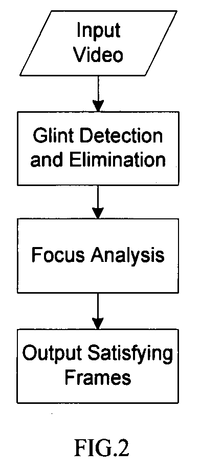
1. System Framework of Automatic Image Quality Assessment

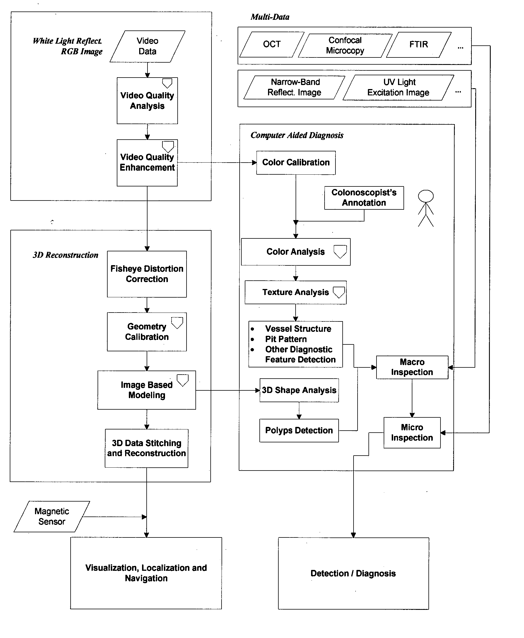
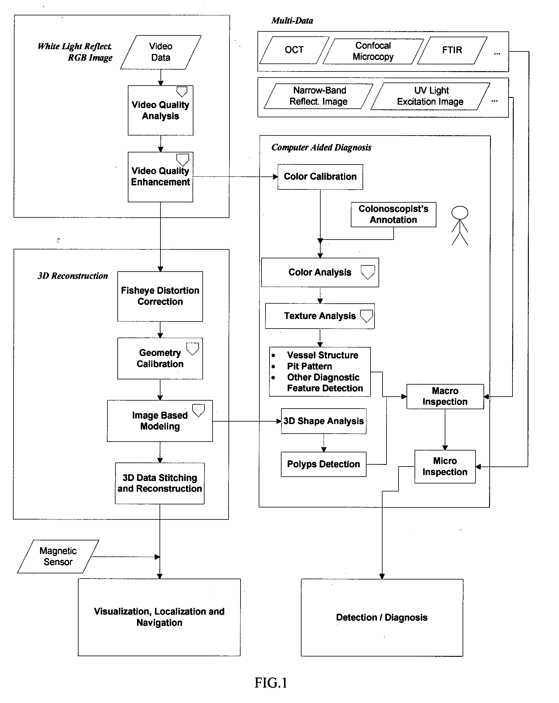
[0029] The present invention is a complex multi-sensor, multi-data and multi-algorithm image processing system. The design provides a modular and open architecture built on phenomenology (feature) based processing. The feature set includes the same features used by the colonoscopists to assess the disease severity (polyp size, pit pattern, etc.). The image-based polyp reconstruction algorithm features several steps: distortion correction, image based modeling, 3D data stitching and reconstruction. The texture-based pit-pattern analysis employs morphological operators to extract the texture pattern, and then utilizes a statistical model and machine learning algorithms to classify the disease severity according to the color and texture information of pits. By analyzing the 3D poly shape and pit-pattern the colonoscopist is provided with diagnostic information for macroscopic inspection. The open architecture also allows for a ...


PUM


Abstract

A process for providing computer aided diagnosis from video data of an organ during an examination with an endoscope, comprising analyzing and enhancing image frames from the video and detecting and diagnosing any lesions in the image frames in real time during the examination. Optionally, the image data can be used to create a 3 dimensional reconstruction of the organ.

Description

[0001] This application claims priority to US provisional patent application 60 / 839275 filed Aug. 21, 2006, incorporated herein by reference.TECHNICAL FIELD [0002] This invention generally relates to medical imaging and more specifically to image processing and computer aided diagnosis for diseases, such as colorectal cancer, using an automated image processing system providing a rapid, inexpensive analysis of video from a standard endoscope, optionally including a 3 dimensional (“3D”) reconstructed view of the organ of interest, such as a patient's colon. This invention is to be used in real time employing video data from a conventional endoscope that is already being used during an examination, such as a colonoscopy, to provide an instantaneous second opinion without substantially prolonging the examination. BACKGROUND ART [0003] Although this invention is being disclosed in connection with colorectal cancer, it is applicable to many other areas of medicine. Colorectal cancer is t...

Claims


Application Information

Patent Timeline
Patent Type & Authority Applications(United States)
IPC IPC(8): A61B1/04
CPCG06T7/0012G06T7/0075G06T2200/08G06T2207/30028G06T5/005G06T2207/20152G06T5/008G06T5/40G06T2207/10016G06T2207/10024G06T2207/20036G06T5/006G06T7/593A61B1/000094
Inventor GU, JIAWOLTERS, ROLF HOLGER
Owner CADES SCHUTTE A LIMITED LIABILITY LAW PARTNERSHIP
Why Eureka
Try Eureka