Such
instability may include, for example, intentional or accidental
tears, cuts, stretching, loosening, deterioration of structure, loss of firmness, and the like.
No matter what type of
instability is present or whether the tissue to be repaired is classified as orthopedic or non-orthopedic, similar issues and objectives are encountered by the surgeon, namely, creating a stable and reliable structure and doing so in as easy and reliable manner as possible.
For example, certain joints, such as hips, knees, shoulders, and elbows contain tissues that are common sources of problems, whether natural or induced, that require extensive
physical therapy or
surgery to correct.
Of such tissues or tissue structures, a
common source of medical problems occurs in the joints.
However, adequate arthroscopic methods that approximate the clinical outcome achieved by open
surgical methods for reducing excessive joint laxity have been slow to develop or have begun to show less than optimal long term clinical outcomes (e.g.
thermal methods).
The
shoulder joint, in particular, has inherent instability because of its
large range of motion combined with the relatively shallow joint bony socket (glenoid).
Damage to or laxity of one of these stabilizing structures can result in the presentation of clinically relevant shoulder instability.
Most typically, the instability of the shoulder stems from disruption and / or looseness (excessive
capsule laxity) of the shoulder
capsule.
The resulting subluxation or
dislocation of the joint can be painful and debilitating for the individual.
A loose shoulder
capsule may be tightened readily when a standard open incision is used, but tightening the shoulder capsule arthroscopically poses significant challenges with existing instruments.
For example, the acute angles at which the surgical devices are able to approximate the soft tissue and identify regions where suturing would be desirable are limiting.
Furthermore, the ability to pass a suture and tie snug surgical knots that compress the tissue in the desired plane with a reasonable suture time is difficult if not cumbersome.
Finally, the ability to dictate the level of tissue tied is limited to the tissue needle bite size and remains difficult for the surgeon to reproducibly specify the level of
tissue compression desired.
It has been demonstrated that at temperatures above 65 degrees Celsius, collagen begins to denature (e.g., unwinding of the
helical structure), resulting in tissue shrinkage.
However, this technology has yielded equivocal results and progressive skepticism from shoulder surgeons.
Additional concerns of thermal capsulorrhaphy application include potential injury to the axillary nerve, bleeding, pain, and excessive swelling of the capsule.
More importantly, the technical methods used during thermal capsulorrhaphy do not allow the surgeon to control the level of plication that is desired or anticipated.
The paucity of data demonstrating the long-term mechanical characteristics and viability of these treated ligaments limits the confident and continued use of this technique.
Conventional methods for arthroscopic plication of the shoulder capsule with sutures typically involve freehand techniques that are technically challenging and often time-consuming.


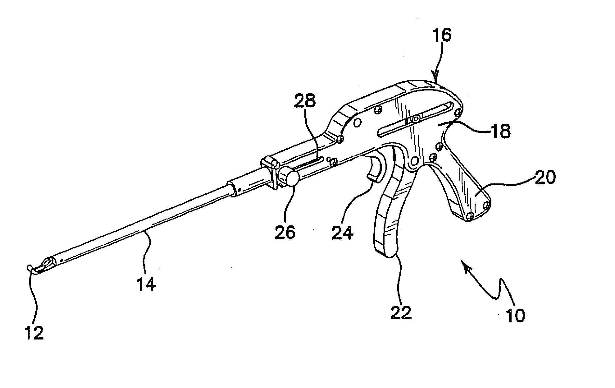
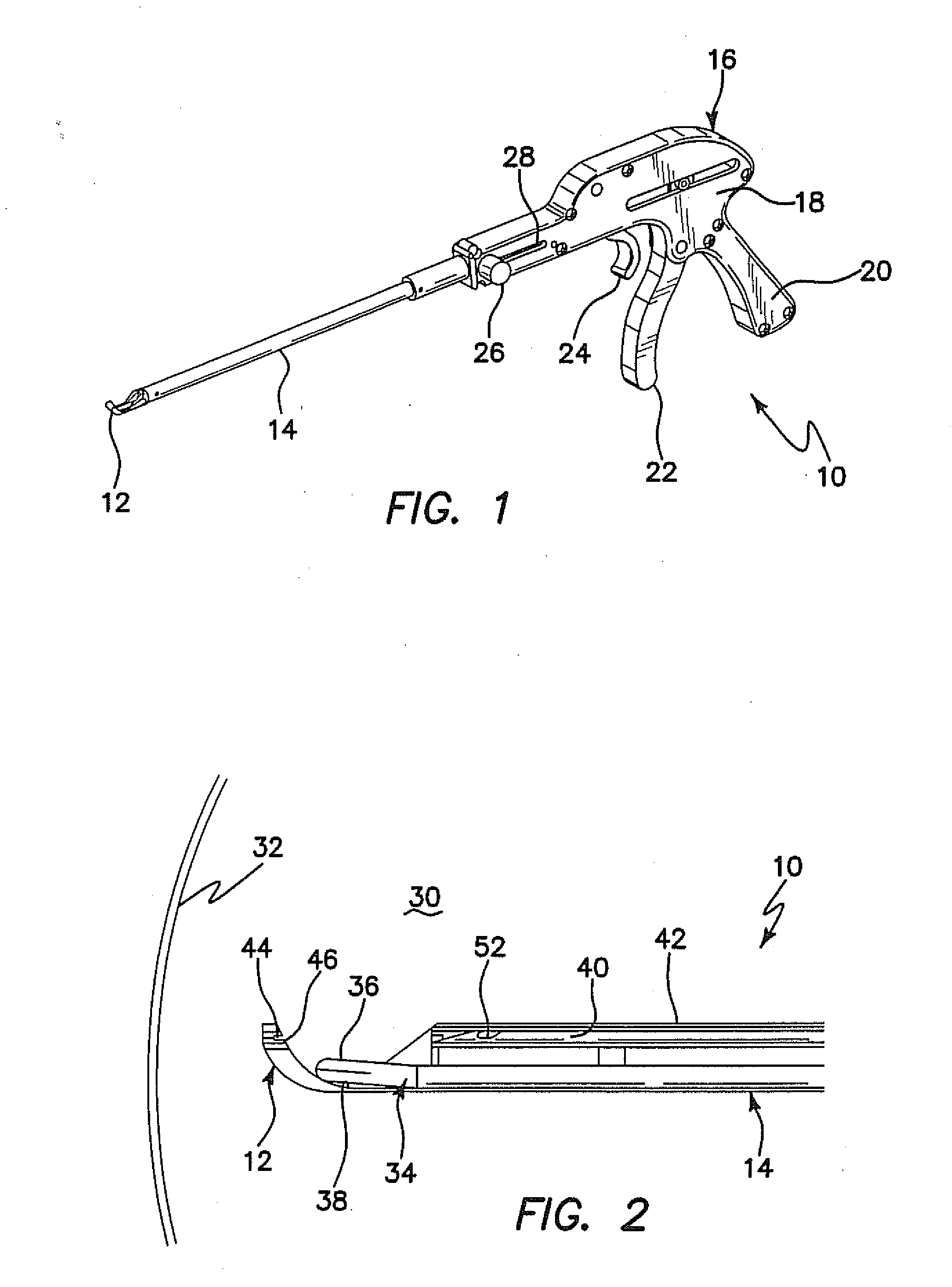
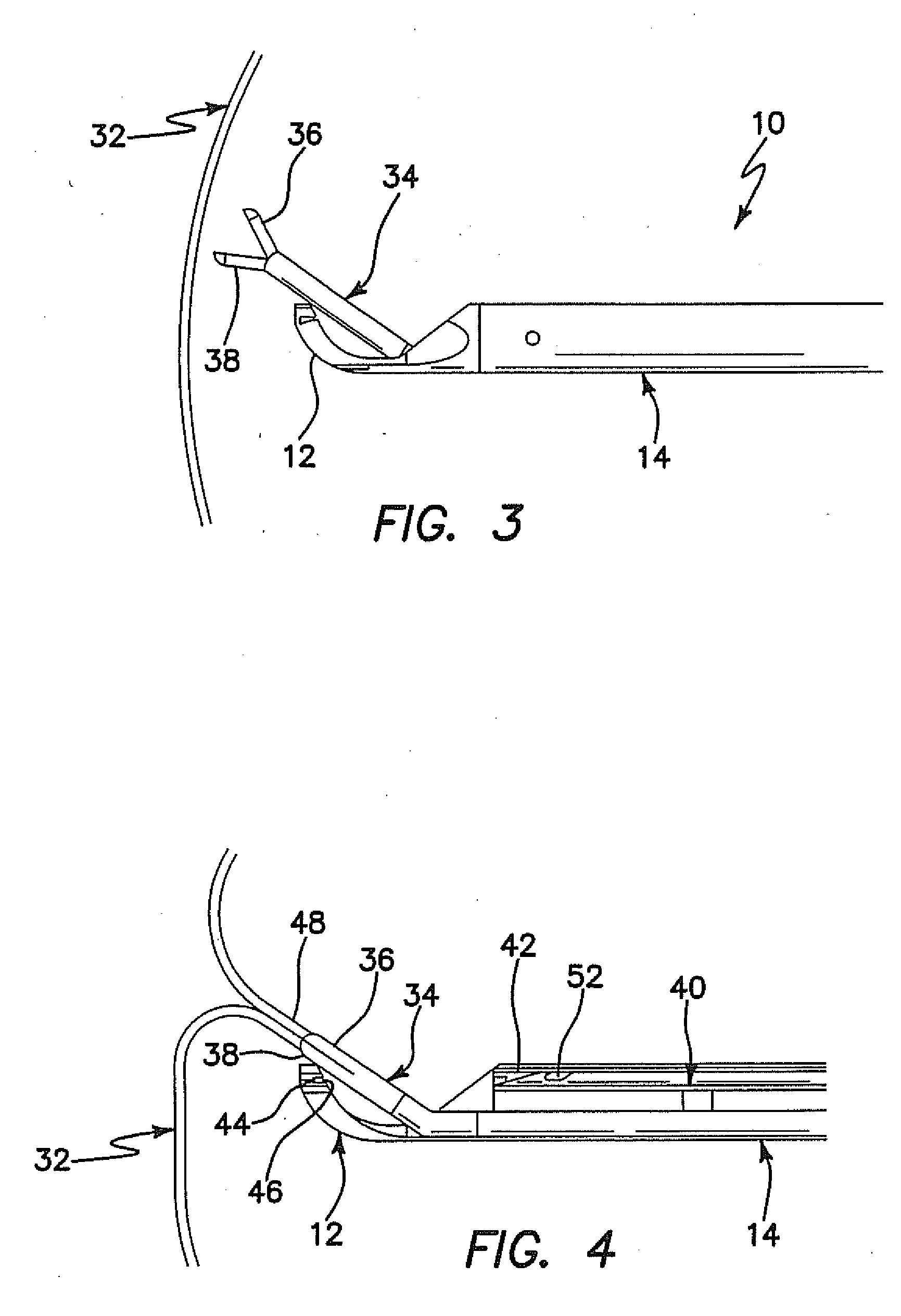