Image Processing System for Automatic Segmentation of a 3-D Tree-Like Tubular Surface of an Object, Using 3-D Deformable Mesh Models

a technology of image processing system and tree-like tubular surface, which is applied in the field of image processing system for automatic segmentation of tree-like tubular surface of object, can solve the problems of difficult mapping of discrete deformation model onto the different branches of tree-like tubular organ, gaps or folds or other deformations at embranchment locations, etc., and achieves the effect of minimizing the number of branch fusion operations

Inactive Publication Date: 2008-04-24
KONINKLIJKE PHILIPS ELECTRONICS NV
View PDF14 Cites 94 Cited by
  • Summary
  • Abstract
  • Description
  • Claims
  • Application Information

AI Technical Summary

Benefits of technology

[0004]The present invention has for an object to propose an image processing system for tree-like tubular structure segmentation. The system of the invention has means for fast tree-like tubular surface mesh generation, comprising automatic branch generation, branch labeling and branch fusing, based on cylindrical surface mesh generation. In particular, said system has processing means for creating and using 2-simplex mesh models or triangular mesh models or any other deformable mesh models.
[0006]The invention has for a further object to propose such a system having processing means to minimize the number of branch fusions. Since the system has means to automatically label the generated tree-like tubular mesh surfaces according to the various branches of the initial tubular tree, the labeling defines various regions of the final tree-like tubular mesh. A first cylindrical structure is generated from the greatest possible number of adjacent centerline segments, in a continuous manner. Then other cylindrical structures are fused to this first cylindrical structure. Creating this first cylindrical structure, which directly forms a main branch from several adjacent centerline segments, to which other branches are fused, minimizes the number of fusions operations. The same principle may be applied to the other branches with sub-branches. Labeling the different regions of the object of interest is of great help while using the mesh as an active model for 3D tree-like organ segmentation in 3-D medical images.

Problems solved by technology

Problems arise when the deformable model, whether of the kind called 2-Simplex Mesh, triangular Mesh or of any other kind of active contour Models, must fit an organ that presents a tree-like tubular structure.
It is very difficult to map the discrete deformable model onto the different branches of the tree-like tubular organ, particularly at the location of the embranchments.
If the merging of the tubular models is not correct, there may be gaps or folds or other deformations at embranchment locations.

Method used


Image

  • Image Processing System for Automatic Segmentation of a 3-D Tree-Like Tubular Surface of an Object, Using 3-D Deformable Mesh Models
  • Image Processing System for Automatic Segmentation of a 3-D Tree-Like Tubular Surface of an Object, Using 3-D Deformable Mesh Models
  • Image Processing System for Automatic Segmentation of a 3-D Tree-Like Tubular Surface of an Object, Using 3-D Deformable Mesh Models

Examples


Embodiment Construction

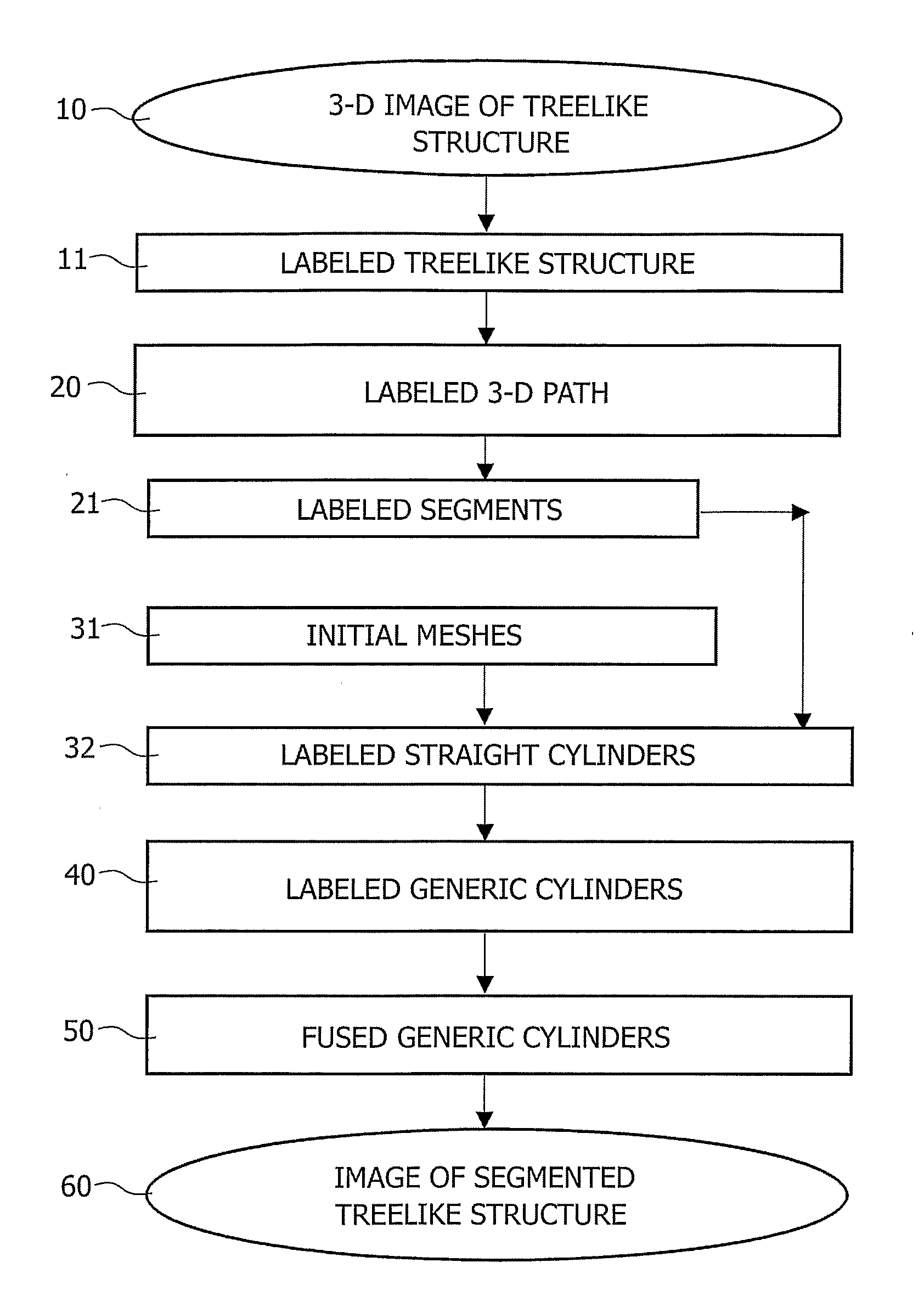
[0018]The invention relates to an image processing system with means of processing three-dimensional (3-D) digital image data. FIG. 1A is a diagrammatic representation of an embodiment of this system. The 3-D image 10 may represent in gray levels the three-dimensional surface of a tubular organ called object of interest OI in a noisy image. In order to provide the user with a better view of the object of interest, for instance with respect to the noisy background, this object is segmented. Segmentation permits the user to better study or detect abnormalities of the organ. The images can be acquired by different acquisition means such as ultrasound or X-ray apparatus or by other apparatus known to those skilled in the art.

[0019]The present invention particularly relates to such an image processing system with means of segmentation of a tree-like tubular object of interest, in a three-dimensional image 10 or in a sequence of three-dimensional images. As illustrated by FIG. 6A, the tre...


PUM


Abstract

An image data processing system with computing means for the automatic segmentation of a treelike tubular structure in a 3-D image comprising: means (20) for computing a treelike center path of the tubular tree-like structure; means (21) for dividing the treelike center path of the tubular treelike structure into segments formed of points; means (40) for generating generic cylindrical meshes formed of cells, for individual segments of the tree-like center path; means (50) for fusing generic cylindrical meshes by two.

Description

FIELD OF THE INVENTION[0001]The invention relates to an image processing system for automatic segmentation of a 3-D tree-like tubular surface of an object in a three-dimensional image, using 3-D deformable mesh models. The invention also relates to a medical examination apparatus using such a system. The invention further relates to program products for processing medical three-dimensional images produced by this apparatus. The invention also relates to a medical image processing method for the segmentation of tubular tree-like body organs such as arteries, for improving the visualization of the organs. The invention finds a particular application in the field of medical imaging.BACKGROUND OF THE INVENTION[0002]A technique of modelization of a 3-D object is already disclosed by H. DELINGETTE in the publication entitled “Simplex Meshes: a General Representation for 3D shape Reconstruction” in the “processing of the International Conference on Computer Vision and Pattern Recognition (...

Claims


Application Information

Patent Timeline
Patent Type & Authority Applications(United States)
IPC IPC(8): G06T15/00G06T5/00G06T17/20
CPCG06K9/0014G06T7/0083G06T7/0089G06T17/20G06T2210/41G06T2207/10116G06T2207/10132G06T2207/30101G06T2200/04G06T7/12G06T7/149G06V20/695
Inventor ROUET, JEAN-MICHELLAFFARGUE, FRANCKFRADKIN, MAXIM
Owner KONINKLIJKE PHILIPS ELECTRONICS NV
Why Eureka
Try Eureka