Method for Segmenting Digital Medical Image

a digital medical image and segmentation technology, applied in image enhancement, image analysis, instruments, etc., can solve the problems of increasing the complexity of the breast cancer diagnosis task, the detection step, and the reliance on the segmentation step, so as to achieve the effect of improving the outcom

Active Publication Date: 2008-05-01
AGFA NV
View PDF5 Cites 59 Cited by
  • Summary
  • Abstract
  • Description
  • Claims
  • Application Information

AI Technical Summary

Benefits of technology

[0025] According to the present invention a Markov Random Field mode is developed and is applied as an extension to th...

Problems solved by technology

The detection step, relying on the segmentation step is generally considered to be a very complex task.
Finally, digital mammography, by its very nature, is inherently characterized by the error intr...

Method used


Image

  • Method for Segmenting Digital Medical Image
  • Method for Segmenting Digital Medical Image
  • Method for Segmenting Digital Medical Image

Examples


Embodiment Construction

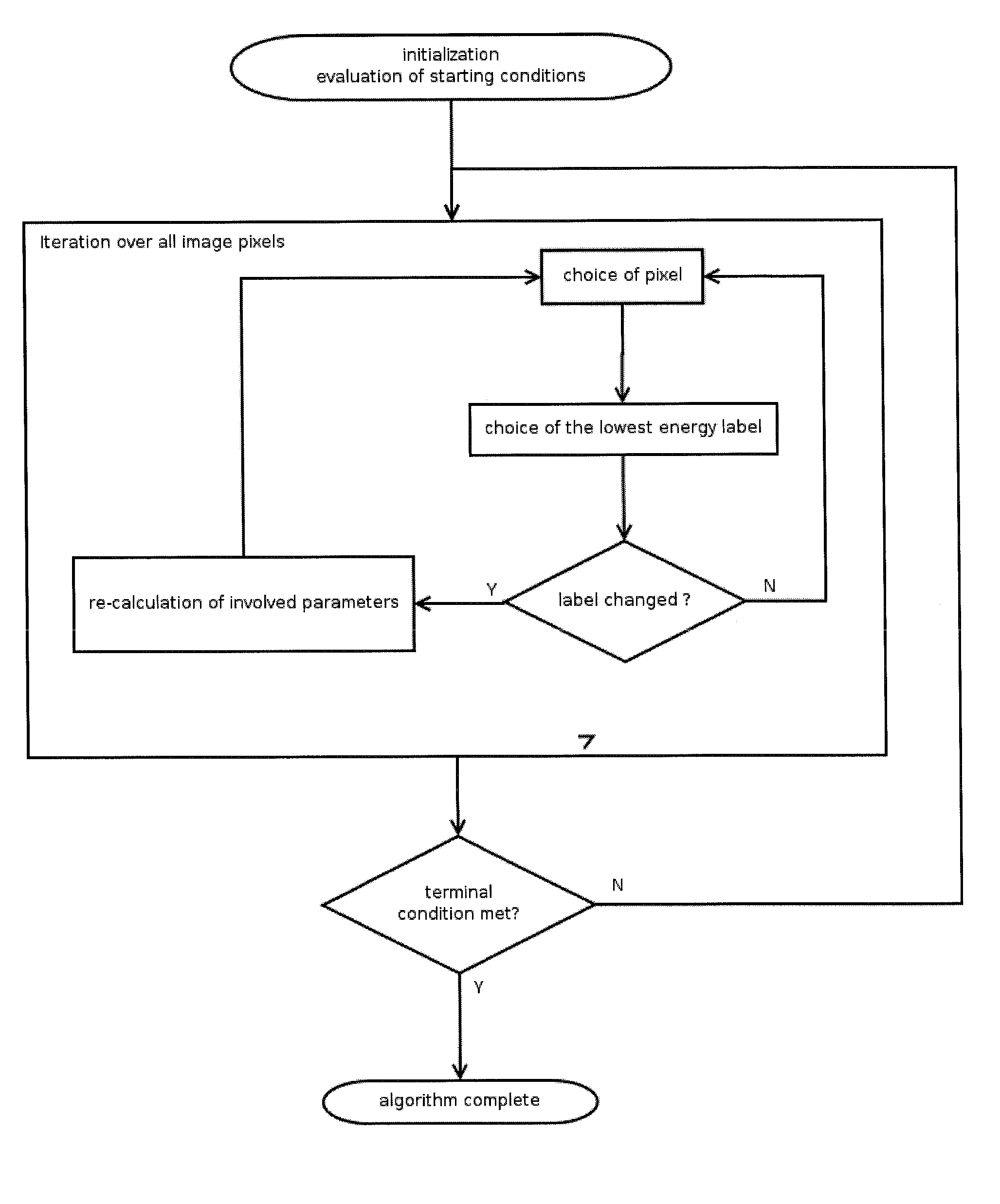
[0046] A new pixel clustering model is disclosed here and applied to the analysis of digital mammograms. The clustering represents the first step in a more general method of mammographic analysis and aims at the creation of a concise data-set (clusters) for automatic detection and classification of mammographic structures such as the pectoral muscle and the breast outline on the one hand and lesions such as masses and micro-calcifications on the other hand. The lesions are typically the first symptoms analyzed in early diagnosis of breast cancer. The image pixels are described by their intensity (grey level) or a multi-dimensional feature vector. Multidimensional decompositions may be obtained by applying multi-scale filter banks or texture operators.

[0047] A Markov Random Field (MRF)-based technique is introduced that is suitable for performing clustering of images characterized by poor or limited data. The proposed method is a statistical classification model that labels the imag...


PUM


Abstract

A Markov Random Field (MRF)-based technique is described for performing clustering of images characterized by poor or limited data. The proposed method is a statistical classification model that labels the image pixels based on the description of their statistical and contextual information. Apart from evaluating the pixel statistics that originate from the definition of the K-means clustering scheme, the model expands the analysis by the description of the spatial dependence between pixels and their labels (context), hence leading to the reduction of the inhomogeneity of the segmentation output with respect to the result of pure K-means clustering.

Description

RELATED APPLICATIONS [0001] This application claims priority to European Patent Application No. EP 06122899.5 filed on Oct. 25, 2006, and claims the benefit under 35 USC 119(e) of U.S. Provisional Application No. 60 / 862,894, filed on Oct. 25, 2006, both of which are incorporated herein by reference in their entirety.BACKGROUND OF THE INVENTION [0002] The anatomy of the female breast changes over age. During the reproductive years, the breast consists mainly of ductal, glandular, and fat tissue. This is interspersed with fibrous tissue providing support and attachment to the chest wall. Glandular and fibrous tissue are jointly called the fibro-glandular tissue. The breast glandular tissue is called the breast parenchyma and consists of 20 . . . 25 lobules (glands), responsible for milk production and are drained towards the nipple by numerous tiny tubes (ducts) that come together to form bigger ducts. Each milk-producing lobule contains a cluster or ring of cells. The sections of lob...

Claims


Application Information

Patent Timeline
IPC IPC(8): G06K9/00G06V10/426
CPCG06K9/4671G06K9/469G06T2207/30068G06T7/0087G06T2207/10116G06T7/0081G06T7/11G06T7/143G06V10/426G06V10/462
Inventor SULIGA, MAREKDEWAELE, PIETDEKLERCK, RUDINYSSEN, EDGARDBEHIELS, GERT
Owner AGFA NV
Why Eureka
Try Eureka