Method for performing distributed analysis and interactive review of medical image data

Inactive Publication Date: 2008-06-19
GENERAL ELECTRIC CO
View PDF3 Cites 19 Cited by
  • Summary
  • Abstract
  • Description
  • Claims
  • Application Information

AI Technical Summary

Benefits of technology

[0007]There is, therefore, significant need for improvement in the proc

Problems solved by technology

Radiology department workflow in a typical hospital or other imaging center, however, requires that many complex analyses be performed on a multitude of different analysis workstations.
A first issue in this typical workflow is the inconvenience cost to radiologists and technicians who essentially access and analyze the image data sequentially, at one or more advanced application workstations.
There is, at present, no convenient mechanism for enabling the radiologist and technician to simply communicate when pre-processing has been completed.
This process can be time consuming and could lend itself to errors in appropriately locating the pre-processed file.
Moreover, the present workflow does not facilitate distributing reports, recommendations or other results of the analysis by radiologists to referring physicians.

Method used


Image

  • Method for performing distributed analysis and interactive review of medical image data
  • Method for performing distributed analysis and interactive review of medical image data
  • Method for performing distributed analysis and interactive review of medical image data

Examples


Embodiment Construction

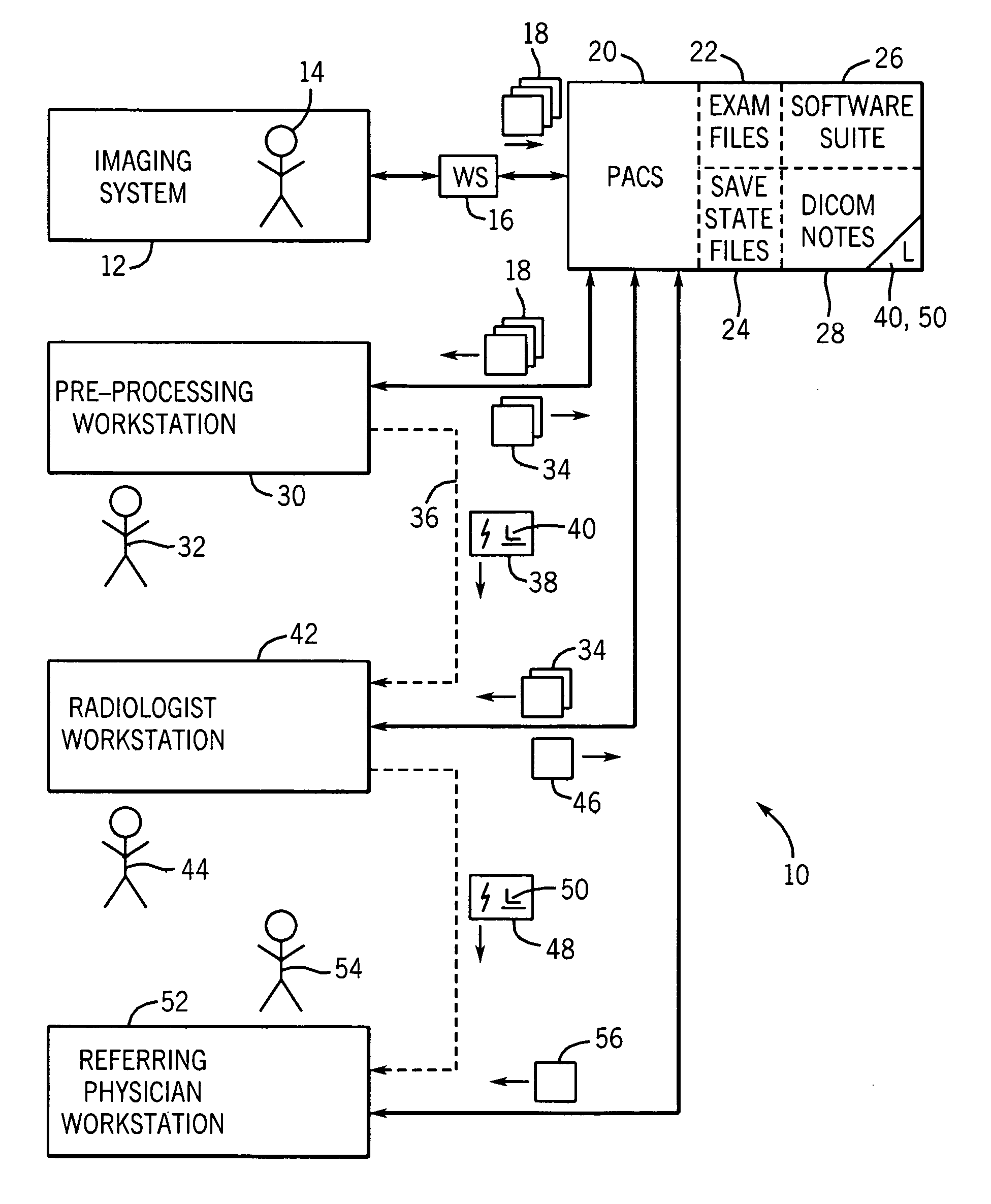
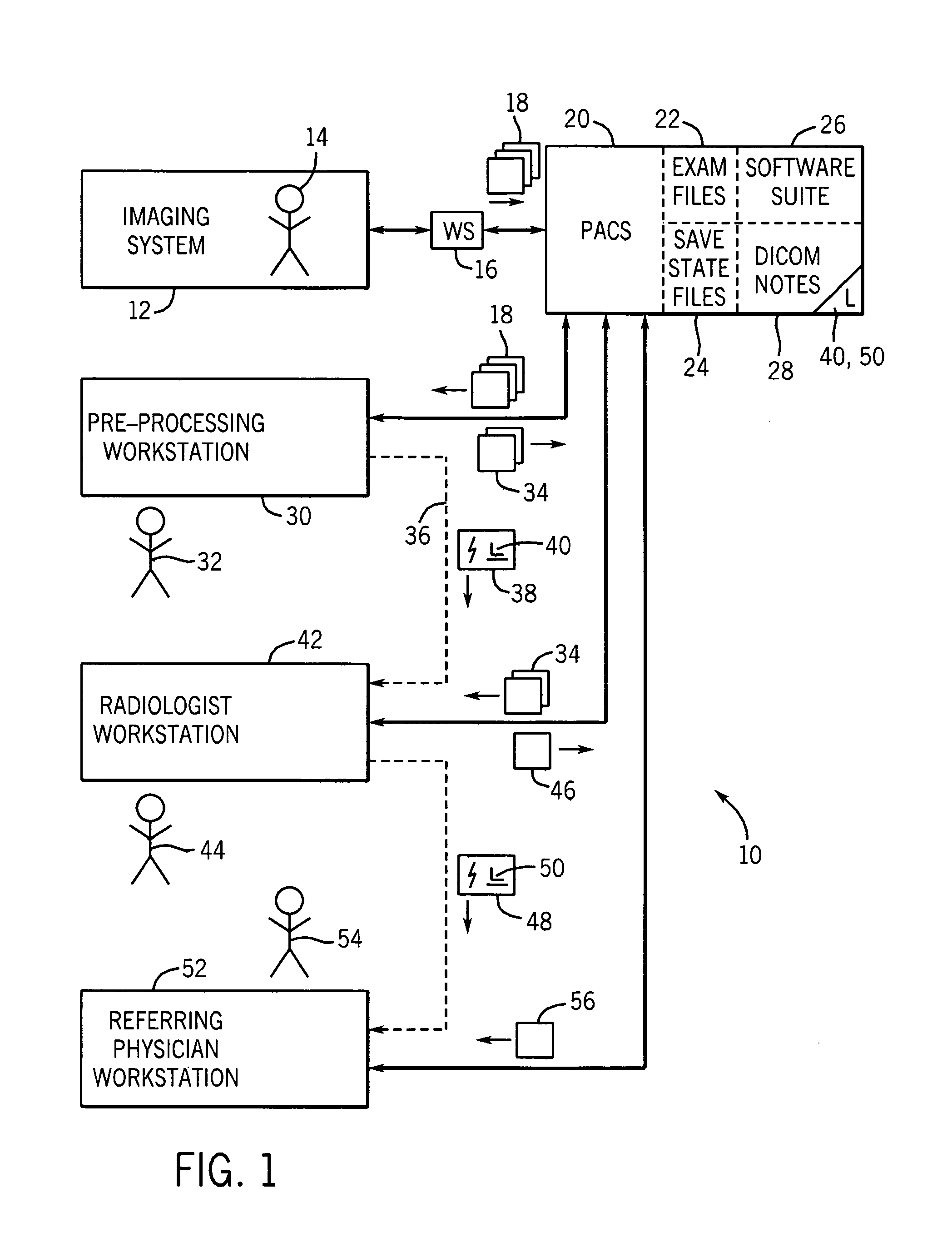
[0019]Turning now to the drawings, and referring first to FIG. 1, an image processing and analysis system, designated generally by reference numeral 10, is illustrated as including an imaging system 12 for generating diagnostic images of a patient 14. As will be appreciated by those skilled in the art, the imaging system 12 may include any current or future modality imaging system capable of making electronic images of anatomies or, more generally, features of interest in the patient. Current technologies envisaged for imaging system 12 include magnetic resonance imaging (MRI), computed tomography imaging (CT), positron emission tomography (PET), ultrasound, X-ray, X-ray tomosynthesis, and so forth, including combinations of these modalities. In general, imaging system 10 will operate under the control of one or more clinicians, technicians or radiologists, who acquire image data by interaction at a workstation 16. The workstation 16 may be wholly or partially integrated with the im...


PUM


Abstract

A technique is provided for improving workflow in processing and exchange of image data in a medical context. Image data may be accessed for processing, and save-state information may be generated following such processing that captures a state of an application used to process the image data files. A link to the save-state information may be stored on a common memory device and selected or executed by a radiologist or downstream service provider for accessing both the images and for placing the application in the state used to process the images. The resulting workflow greatly facilitates access to images as well as restoration of useful states of applications used to process the images.

Description

BACKGROUND[0001]The present invention relates generally to the field of medical image processing. More particularly, the invention relates to techniques for accessing, pre-processing, and finally processing medical images, such as through reads by radiologists and other medical professionals.[0002]In a field of medical diagnostic imaging, many processes are known for generating images and for managing their distribution and use. For example, a number of imaging modalities are currently available, such as magnetic resonance imaging (MRI), computed tomography (CT), positron emission tomography (PET), ultrasound, X-ray, X-ray tomosynthesis, and so forth. In general, patients undergo such examinations to identify anatomical structures and to enable medical professionals to evaluate the anatomical features and functions for health and possible disease states, as well as response to treatment, and so forth.[0003]In a common scenario, digital information is gathered from one of the imaging...

Claims


Application Information

Patent Timeline
IPC IPC(8): G06K9/00
CPCG06F19/321G16H30/20G16H50/20G16H80/00
Inventor LAL, RAKESH MOHANSIROHEY, SAAD AHMEDPATZ, TABB ALAN
Owner GENERAL ELECTRIC CO
Why Eureka
Try Eureka