Intravascular implantable device having detachable tether arrangement

Inactive Publication Date: 2008-06-19
SYNECOR LLC
View PDF55 Cites 42 Cited by
  • Summary
  • Abstract
  • Description
  • Claims
  • Application Information

AI Technical Summary

Benefits of technology

[0012]In one embodiment, the vascular anchor is separate from the implantable device and captures a tether portion that extends from the implantable device between the anchor and the vasculature. In another embodiment, the anchor may be incorporated as part of the implantable device. In one embodiment, the vascular anchor and/or the implantable device include mechanisms to optimize interference between the anchor and the tether portion in a way that does not induce a rupture of the vessel while providing for adequate clinical attachment of the implantable device within the patient.
[0013]Unlike the previous approaches to extracting an intravascular implantable device anchored near the middle of the patient's torso, embodiments of the present invention utilize a detachable tether arrangement positioned proximate and end of the elongated body portion of the intravascular impla

Problems solved by technology

A secondary, fallback access that can occur by a subclavian vessel puncture in the event that th

Method used


Image

  • Intravascular implantable device having detachable tether arrangement
  • Intravascular implantable device having detachable tether arrangement
  • Intravascular implantable device having detachable tether arrangement

Examples


Embodiment Construction

[0048]In the following detailed description of the present invention, numerous specific details are set forth in order to provide a thorough understanding of the present invention. However, one skilled in the art will recognize that the present invention may be practiced without these specific details. In other instances, well-known methods, procedures, and components have not been described in detail so as to not unnecessarily obscure aspects of the present invention.

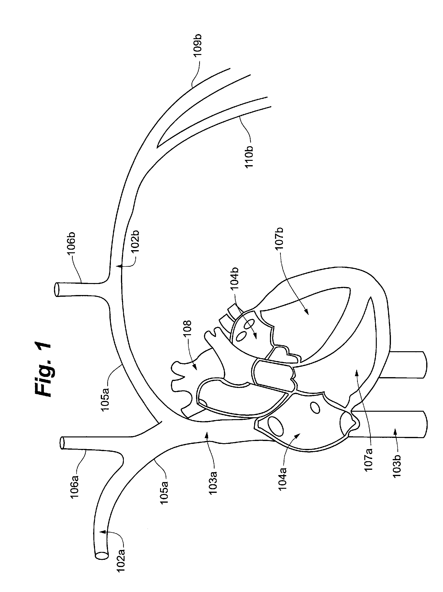
[0049]Referring now to FIG. 1, the general cardiac anatomy of a human is depicted, including the heart and major vessels. The following anatomic locations are shown and identified by the listed reference numerals: Right Subclavian 102a, Left Subclavian 102b, Superior Vena Cava (SVC) 103a, Inferior Vena Cava (IVC) 103b, Right Atrium (RA) 104a, Left Atrium (LA) 104b, Right Innominate / Brachiocephalic Vein 105a, Left Innominate / Brachiocephalic Vein 105b, Right Internal Jugular Vein 106a, Left Internal Jugular Vein 106b, Ri...


PUM


Abstract

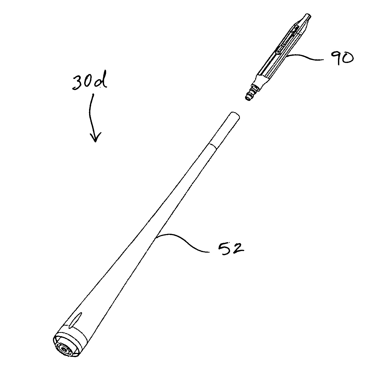
An intravascular implantable medical device includes a detachable tether arrangement. In one embodiment, the detachable tether arrangement comprises a tip assembly that includes a break-away joint and a tether portion, and may also include a connector and an extension portion connected to an end of an elongated intravascular implantable medical device. In this embodiment, the break-away joint is disposed between the extension portion and the tether portion. In some embodiments, the extension portion may comprise a housing or other arrangement that includes an antenna for transmitting and receiving data. In other embodiments, the extension portion may also include a lead for defibrillation, pacing, and/or sensing cardiac electrical activity. In certain embodiments, the tether arrangement is designed to be secured within a patient's vasculature with a vascular anchor either in the form of a separate anchor, such as a conventional stent, or by various integrated retention arrangements.

Description

RELATED APPLICATIONS[0001]The present application claims the benefit of U.S. Provisional Application No. 60 / 868,434, filed Dec. 4, 2006, and U.S. Provisional Application No. 60 / 868,437, filed Dec. 4, 2006, and U.S. Provisional Application titled “Implantation Methods, Systems and Tools for Intravascular Implantable Devices”, filed Dec. 3, 2007, the disclosures of which are hereby incorporated by reference in their entireties.FIELD OF THE INVENTION[0002]The present invention relates generally to surgical devices and methods for retaining medical devices within the body, and more specifically to a detachable tether arrangement for a medical device and a method of extracting an intravascular implantable medical device from within a vessel.BACKGROUND OF THE INVENTION[0003]Implantable medical devices such as pacemakers, defibrillators, and implantable cardioverter defibrillators (“ICDs”) have been successfully implanted in patients for years for treatment of heart rhythm conditions. Pace...

Claims


Application Information

Patent Timeline
IPC IPC(8): A61F2/06A61F2/82
CPCA61F2/82A61N1/375A61N1/0563A61N1/056A61N1/37512A61N1/37516A61N1/37518
Inventor RANSBURY, TERRANCEHOLBROOK, KEVIN
Owner SYNECOR LLC
Why Eureka
Try Eureka