Method for image registration processes and X-ray angiography system

a technology of image registration and x-ray angiography, applied in the field of image registration processes and x-ray angiography systems, can solve the problems of insufficient information, difficult to recognize on x-ray images, and insufficient patient data, and achieve the effect of expanding the possibilities for patient data

Active Publication Date: 2008-09-04
SIEMENS HEALTHCARE GMBH
View PDF13 Cites 15 Cited by
  • Summary
  • Abstract
  • Description
  • Claims
  • Application Information

AI Technical Summary

Benefits of technology

[0007]The object of the invention is therefore to extend the possibilities for usi

Problems solved by technology

During an operation, additional images are then acquired which in themselves do not necessarily provide sufficient information.
Although soft parts can be well imaged by MRI, they are difficult to recognize on X-ray images.
In the case of MRI, however, the image volume which can be acquired within typical acquisition times of 20 sec

Method used


Image

  • Method for image registration processes and X-ray angiography system
  • Method for image registration processes and X-ray angiography system

Examples


Embodiment Construction

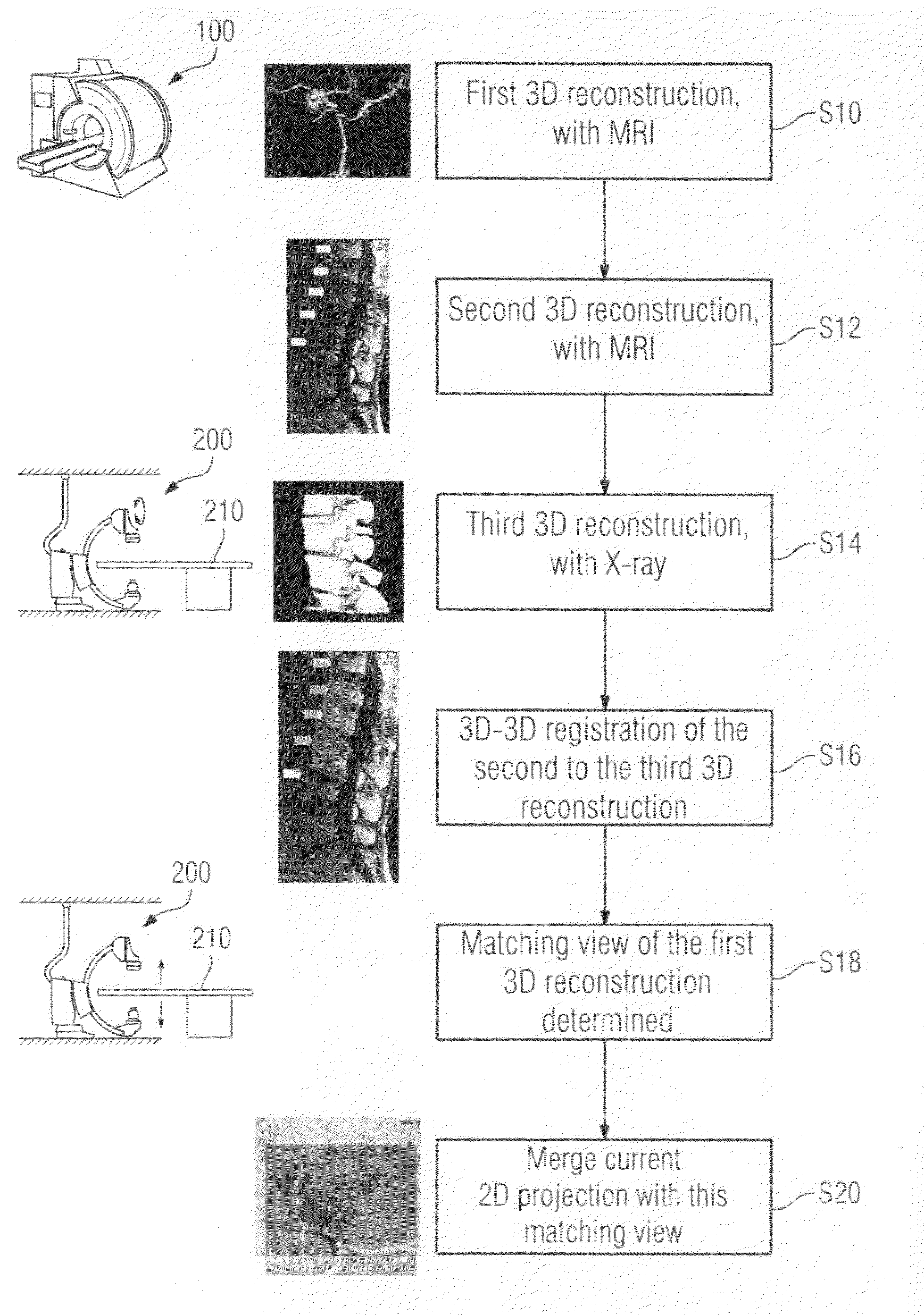
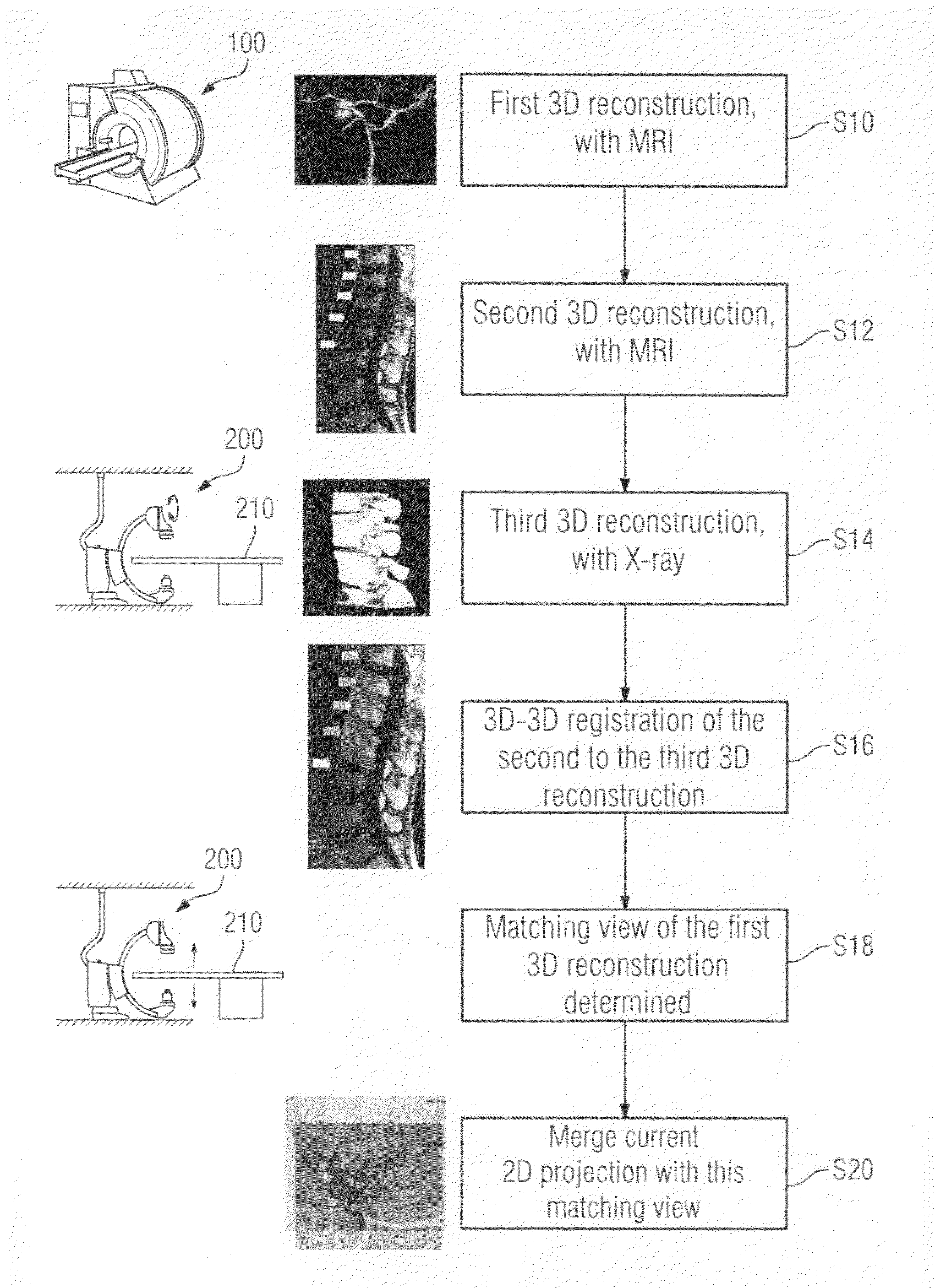
[0032]In a patient's body there is an area of interest to the treating doctor which in the present case shall be located in a soft part. For example, the doctor is interested in the left atrium of the heart in order to prepare an atrial ablation. An MRI scanner 100 is optimally suited for visualizing such soft parts. In step S10 of the method according to the invention, a sequence of 2D slices of the area of interest is accordingly acquired preoperatively, the image acquisition parameters being optimally selected for imaging the area of interest and there being obtained in known manner from the sequence of 2D slices a 3D reconstruction which, in the present case, constitutes a first 3D image data set. Directly thereafter, with the patient's position in the MRI scanner 100 remaining unchanged, a sequence of 2D slices of vertebral bodies is acquired, this time the image acquisition parameters creating optimum conditions for good visualization of vertebral bodies, and there being gener...


PUM


Abstract

Registration of preoperatively acquired MRI images of soft parts to intraoperatively acquired X-ray images of soft parts is not possible. The invention shows a way of nevertheless using such preoperatively acquired images for superimposition with 2D projections of the soft parts, taking an indirect route via 3D/3D registration of images of the spinal column. For this purpose, 3D image data sets of the spinal column must be obtained separately on the one hand using MRI and on the other using the X-ray imaging system so that the 3D/3D registration produces a mapping rule which then also applies to the preoperatively acquired images of the soft part if the soft part images and the spinal images are acquired without intervening change in the patient position in the MRI scanner.

Description

CROSS REFERENCE TO RELATED APPLICATIONS[0001]This application claims priority of German application No. 10 2007 010 806.2 filed Mar. 2, 2007, which is incorporated by reference herein in its entirety.FIELD OF THE INVENTION[0002]The invention relates to a method for creating extended possibilities for using patient image data that is unsuitable for the application of registration processes, and an X-ray angiography system for use in conjunction with the inventive method.BACKGROUND OF THE INVENTION[0003]These are registration processes which create extensive imaging possibilities to assist a treating doctor during a medical intervention or surgical procedure. Image data sets are regularly acquired preoperatively using image acquisition systems which show the image area of interest in an optimum manner. During an operation, additional images are then acquired which in themselves do not necessarily provide sufficient information. The previously acquired image data is now registered to t...

Claims


Application Information

Patent Timeline
IPC IPC(8): G06K9/00
CPCA61B2019/5289G06T7/0038G06T2207/10072G06T2207/30048G06T2207/10088G06T2207/30004G06T2207/30012G06T2207/10081A61B2090/364G06T7/38
Inventor BOESE, JANJUSTIZ, JORNMEYER, ANDREASPFISTER, MARCUS
Owner SIEMENS HEALTHCARE GMBH
Why Eureka
Try Eureka