Animal model for prostatic stromal hyperplasia

Inactive Publication Date: 2008-10-16
TAIHO PHARMA CO LTD
View PDF0 Cites 0 Cited by
  • Summary
  • Abstract
  • Description
  • Claims
  • Application Information

AI Technical Summary

Benefits of technology

[0057](c) selecting a test substance that reduces the stromal area ratio or weight based on the result obtained in (b) in comparison with the stromal area ratio or weight of the implanted tissue of the non-human animal model for prostatic stromal hyperplasia before administering the test substance.

Problems solved by technology

However, the matured prostate glands derived from the mouse and rat obtained by the above-described method exhibited epithelial-dominant tissue structures, which are different from the stroma-dominant tissue structures observed in human benign prostatic hyperplasia.

Method used


Image

  • Animal model for prostatic stromal hyperplasia
  • Animal model for prostatic stromal hyperplasia
  • Animal model for prostatic stromal hyperplasia

Examples


example 1

Production of Animal Model having Prostate-Like Stroma-Enlarged Tissue

(1) Removal of Rat Fetal Urogenital Sinus

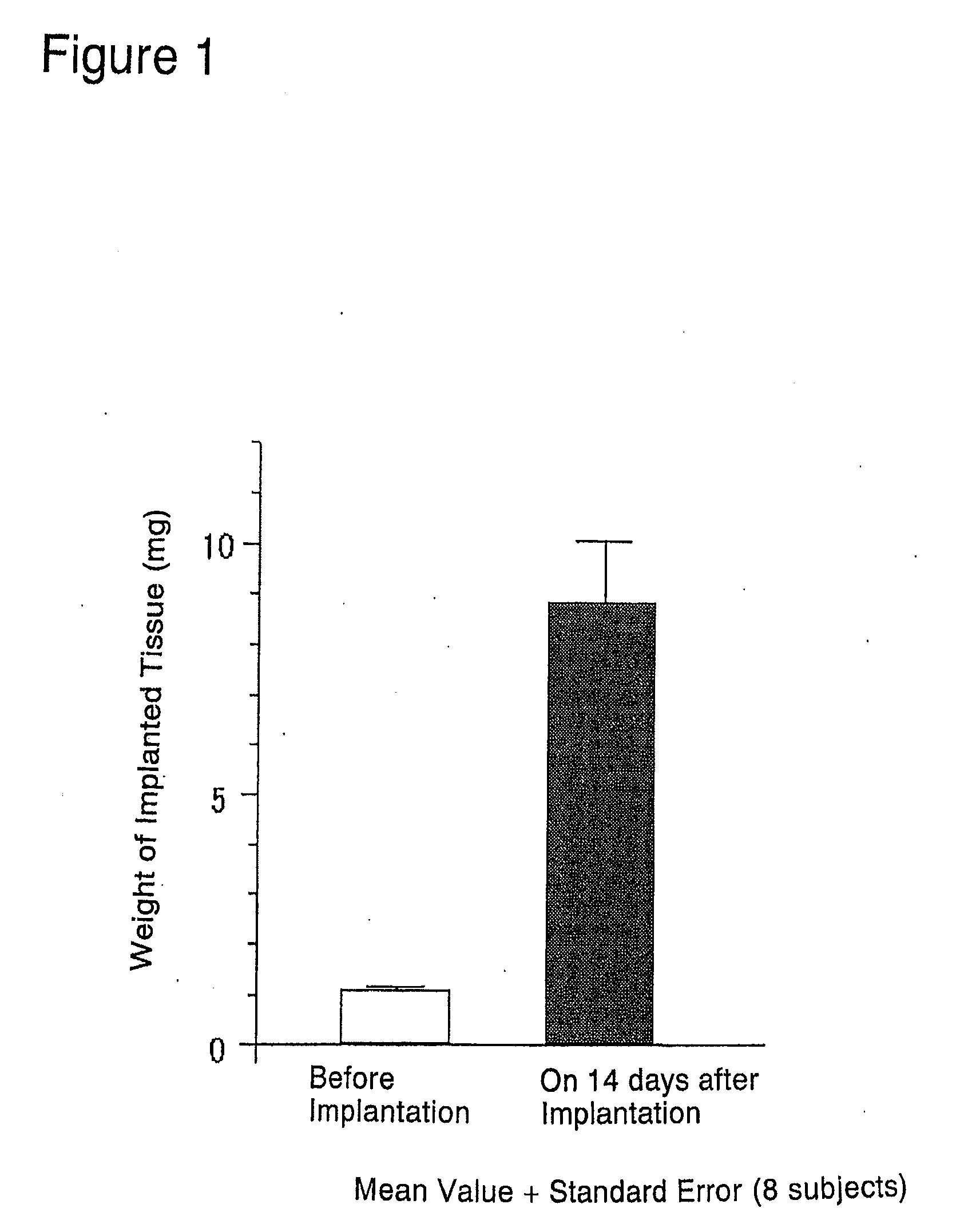
[0171]Under ether anesthesia, fetuses were removed from female SD rats that were 21 days pregnant. From the removed male fetuses, urogenital sinuses were removed also under ether anesthesia while observing through a stereoscopic microscope. The urogenital sinuses were stored in a sterilized culture medium until implantation.

(2) Implantation Procedure

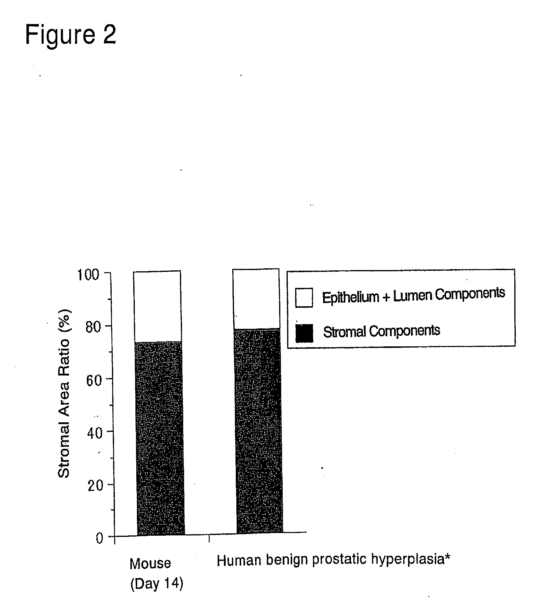
[0172]The abdominal skin adjacent the left femoral region of 6-week old BALB / c male nude mice was cut open about 2 to 3 mm under ether anesthesia. A single rat fetal urogenital sinus obtained in (1) above was subcutaneously implanted after incision using a forceps and the incision was sutured. Thereafter, these nude mice were reared in clean racks (Rat Bracket Cages, 3 to 4 mice / cage) for 2 weeks under the following conditions:[0173]Temperature: 20 to 26° C.[0174]Humidity: 30 to 70%[0175]Lighting hours: 12-hour light-dark cycle;...

example 2

Production of Animal Model for Prostatic Stromal Hyperplasia

(1) Removal of Rat Fetal Urogenital Sinus

[0186]Under ether anesthesia, fetuses were removed from female SD rats that were 20 days pregnant. From the removed fetuses, the urogenital sinuses were removed under ether anesthesia while observing through a stereoscopic microscope. The urogenital sinuses were stored in a sterilized culture medium until implantation.

(2) Implantation Procedure

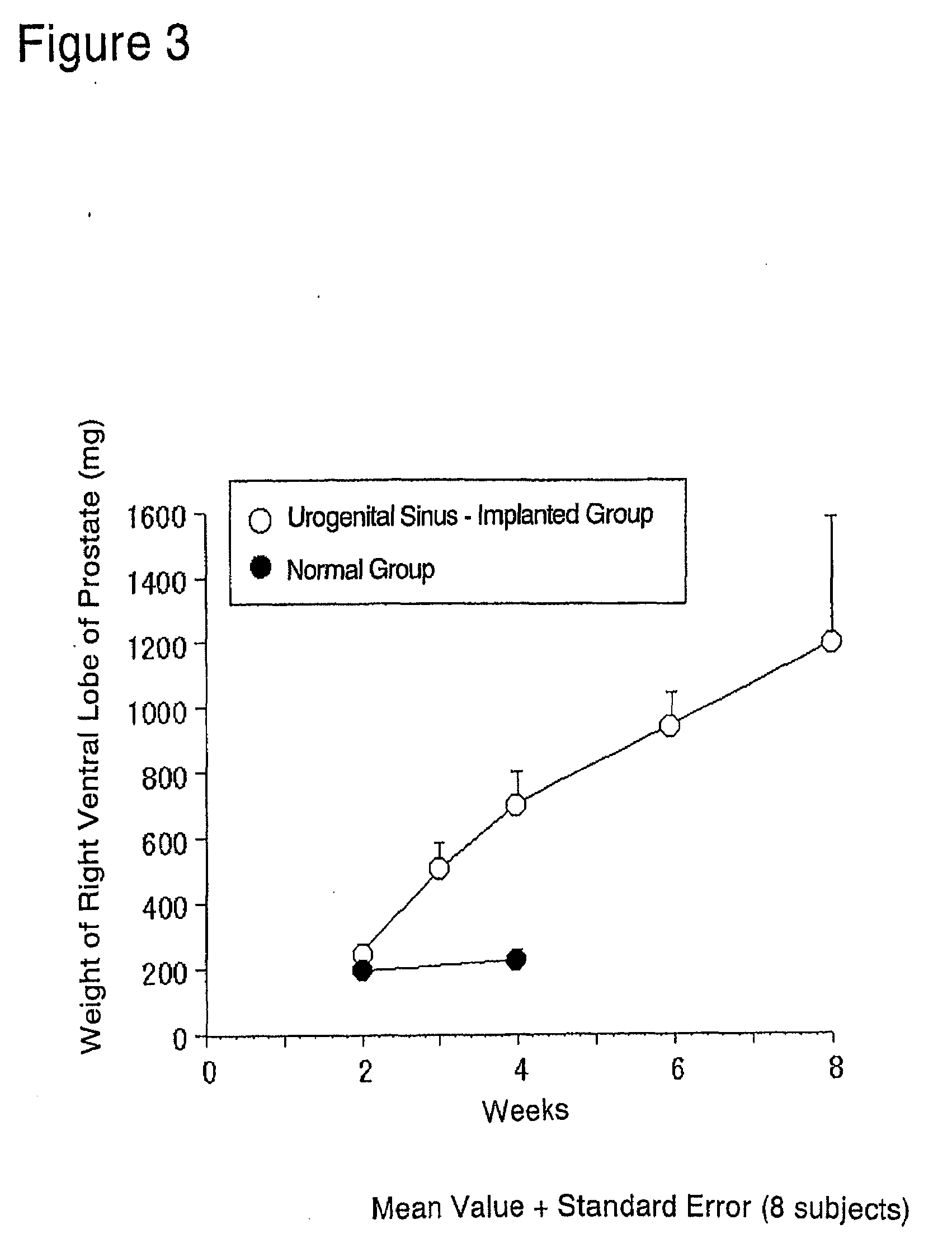
[0187]The prostate glands were exposed by midline incision using 6-week old male SD rats under ether anesthesia. The capsule of prostate right ventral lobe was slightly cut open while observing through a stereoscopic microscope. Two of the fetal urogenital sinuses obtained in (1) above were implanted using a forceps beneath the capsule, and the incision in the capsule was sutured. The cut abdominal skin then closed and the rats were reared for 2 to 8 weeks in normal rearing cages (Rat Bracket Cages, 3 to 4 rats / cage) under the following conditi...

example 3

Production of Animal Model for Prostatic Stromal Hyperplasia

(1) Removal of Rat Fetal Urogenital Sinus

[0201]Urogenital sinuses were removed from the fetuses of female SD rats (donor animal) that were 20 days pregnant in the same manner as in Example 2 (1). The urogenital sinuses were stored in a sterilized culture medium until implantation.

(2) Implantation Procedure

[0202]In the same manner as in Example 2 (2), a single rat fetal urogenital sinus obtained in (1) above was implanted beneath the capsule of prostate right ventral lobe of each 6-week old male SD rat (recipient animal) (n=18×2), and the prostatic capsule was sutured. The implanted rats were reared in normal rearing cages for 3 to 6 weeks thereafter.

(3) Weighing of Implanted Tissue (Rat Fetal Urogenital Sinus-Derived Tissue)

[0203]The implanted tissues (rat fetal urogenital sinus-derived tissues) were removed from the above-prepared rats under ether anesthesia on 3 weeks (n=18) and 6 weeks (n=18) after implantation, and were...


PUM

PropertyMeasurementUnit
weightaaaaaaaaaa
temperatureaaaaaaaaaa
weightaaaaaaaaaa

Abstract

The present invention provides an animal model for prostatic stromal hyperplasia, and a method for screening for a substance effective for preventing / treating human benign prostatic hyperplasia using the animal model. The animal model for prostatic stromal hyperplasia is produced by implanting the fetal urogenital sinus of a non-human animal under the skin or beneath the prostatic capsule of a non-human animal belonging to the species of the same as or different from the said animal. A substance effective for preventing / treating human benign prostatic hyperplasia can be screened by administering a test substance to the animal model and measuring the preventive or therapeutic effect of the test substance upon the implanted tissue (fetal urogenital sinus or tissue derived therefrom).

Description

CROSS-REFERENCE TO RELATED APPLICATIONS[0001]This application is a divisional of application Ser. No. 10 / 477,077, filed Nov. 7, 2003, which is the U.S. National Phase under 35 U.S.C. §371 of International Application No. PCT / JP02 / 04477, filed May 8, 2002 designating the U.S., which claims priority to Japanese Patent Application JP 2002-138123, filed May 9, 2001, the entire disclosures of which are incorporated herein by reference in their entireties.BACKGROUND OF THE INVENTION[0002]1. Field of the Invention[0003]The present invention relates to an animal model for prostatic stromal hyperplasia and a process for producing it. The invention also relates to a method for screening for a substance effective for preventing or treating human benign prostatic hyperplasia using such an animal model. The invention is further directed to a method for evaluating the preventive or therapeutic effect of each test substance on human benign prostatic hyperplasia.[0004]2. Description of the Related ...

Claims


Application Information

Patent Timeline
Patent Type & Authority Applications(United States)
IPC IPC(8): A61K35/00A01K67/027C12N15/01A61P43/00G01N33/53C12N5/071G01N33/50G01N33/574G01N33/68
CPCA01K67/0271A01K2227/105A01K2267/0331C12N5/0683G01N33/6893G01N2800/342A61P13/08A61P43/00
Inventor ODA, NOBUYUKIMIYOSHI, KAZUHISAHARUNO, AKIHIROMIYAKE, HIDEKAZU
Owner TAIHO PHARMA CO LTD
Why Eureka
Try Eureka