X-ray tomographic imaging apparatus

a tomographic imaging and x-ray technology, applied in tomography, instruments, applications, etc., can solve the problems of inaccurate ratios of pixel values, inability to always be constant, and inability to accurately perceive the diagnosis effect, and accurate weighting factors

Active Publication Date: 2008-10-23
GE MEDICAL SYST GLOBAL TECH CO LLC
View PDF20 Cites 43 Cited by
  • Summary
  • Abstract
  • Description
  • Claims
  • Application Information

AI Technical Summary

Benefits of technology

[0028]According to the present invention, an operator, who makes diagnosis while observing a dual-energy image, can know an accurate weighting factor required to obtain a dual-energy image. Thus,

Problems solved by technology

The weighting factor for use in weighted subtraction processing for obtaining an image of bones or soft tissue, however, is not always constant because of a difference in size among subjects and a difference in X-ray absorption coefficient due to individual differences among subjects.
If diagnosis is made with a dual

Method used


Image

  • X-ray tomographic imaging apparatus
  • X-ray tomographic imaging apparatus
  • X-ray tomographic imaging apparatus

Examples


Embodiment Construction

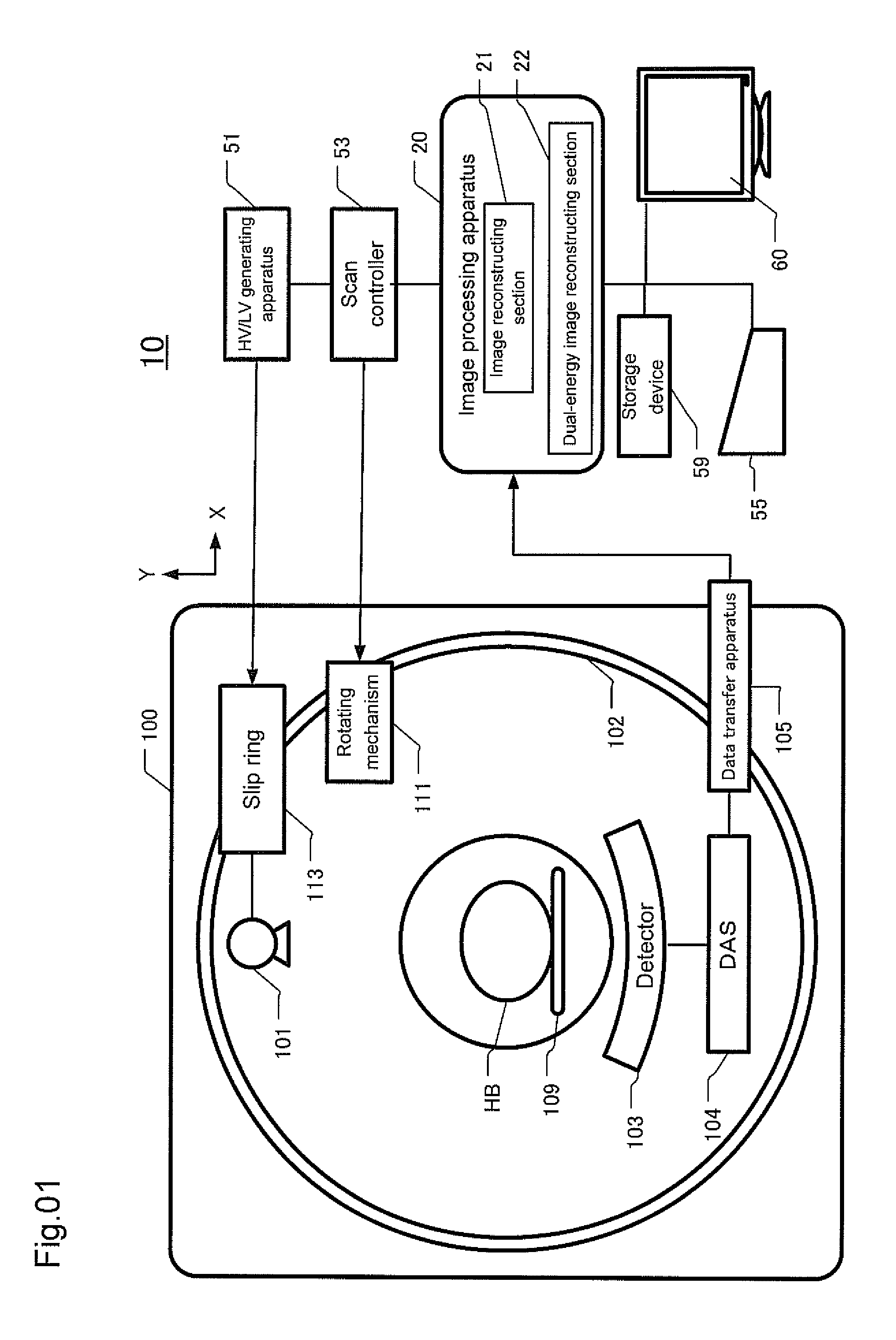
Configuration of an X-Ray Tomographic Imaging Apparatus.

[0039]FIG. 1 is a block diagram showing the configuration of an X-ray tomographic imaging apparatus (X-ray CT apparatus) 10 in accordance with an embodiment of the present invention. The X-ray tomographic imaging apparatus 10 is provided with a gantry 100, and a table 109 for inserting a subject HB into an imaging region in the gantry 100. The table 109 is moved in a Z-direction, which is a direction of a body axis of the subject HB. The gantry 100 has a rotating ring 102, which comprises an X-ray tube 101 for emitting X-rays of a cone beam shape and a multi-row X-ray detector 103 disposed to face the X-ray tube 101. The X-ray tube 101 is configured to emit X-rays having a high-energy spectrum and those having a low-energy spectrum. The multi-row X-ray detector 103 detects X-rays passing through the subject HB.

[0040]The multi-row X-ray detector 103 is formed of scintillators and photodiodes. The multi-row X-ray detector 103 is ...


PUM


Abstract

For the purpose of providing an X-ray tomographic imaging apparatus for displaying a dual-energy image to facilitate diagnosis by an operator, an X-ray tomographic imaging apparatus (10) comprises: an image comparison information calculating section (24) for calculating image comparison information between said first-energy projection dataset (LD) or first-energy tomographic image (LT) and said second-energy projection dataset (HD) or second-energy tomographic image (HT); a region-of-interest defining section (23) for defining a region of interest; a weighting factor determining section (25-2) for determining a weighting factor for use in weighted subtraction processing between said first-energy projection dataset or first-energy tomographic image and said second-energy projection dataset or second-energy tomographic image, such that said image comparison information in said region of interest can be substantially eliminated by conducting said weighted subtraction processing; and a dual-energy image reconstructing section (22) for reconstructing a dual-energy image by conducting weighted subtraction processing between said first-energy projection dataset or first-energy tomographic image and said second-energy projection dataset or second-energy tomographic image used in said image comparison information calculating section, using a weighting factor determined at said weighting factor determining section.

Description

CROSS REFERENCE TO RELATED APPLICATIONS[0001]This application claims the benefit of Japanese Patent Application No. 2006-316030 filed Nov. 22, 2006.BACKGROUND OF THE INVENTION[0002]The present invention relates to an X-ray tomographic imaging technology, and particularly to an X-ray tomographic imaging apparatus pertinent to dual-energy imaging.[0003]In dual-energy imaging, an image can be acquired with a specific material within a subject to be examined highlighted based on X-ray projection data for the imaged subject acquired with X-rays having two different energy spectra. Specifically, an image acquired with high-energy X-rays and that acquired with low-energy X-rays are subjected to addition / subtraction processing, whereby a dual-energy image clearly showing bones and calcified lesions or that clearly showing soft tissue can be obtained.[0004]In performing dual-energy imaging, the projection data acquired with high-energy X-rays and those acquired with low-energy X-rays are obt...

Claims


Application Information

Patent Timeline
IPC IPC(8): H05G1/60G06K9/00
CPCA61B6/032A61B6/4035A61B6/4042A61B6/405A61B6/482A61B6/505G06T11/008A61B6/027
Inventor IMAI, YASUHIRONISHIDE, AKIHIKO
Owner GE MEDICAL SYST GLOBAL TECH CO LLC
Why Eureka
Try Eureka