Medical devices, systems and methods for closing perforations

a technology of perforation and medical devices, applied in the field of tissue perforation closure, can solve the problems of uneven spacing of the anchors around the perforation, difficult to ensure proper approximation of the tissue around the perforation, and difficult to ensure the closure complete, so as to achieve the effect of easy control

Active Publication Date: 2008-11-27
COOK MEDICAL TECH LLC
View PDF104 Cites 78 Cited by
  • Summary
  • Abstract
  • Description
  • Claims
  • Application Information

AI Technical Summary

Benefits of technology

[0010]According to more detailed aspects of the method, the plurality of delivery devices are retracted into the plurality of tubes. A cutting instrument may be passed through an accessory channel of the endoscope and used to form an opening in the bodily tissue. The step of forming the opening is preferably performed after the step of positioning the plurality of delivery devices proximate the bodily tissue, and most preferably after the step of delivering the plurality of tissue fixation devices. The method also includes adjusting the position of the medical system relative to the bodily tissue prior to the step of passing the plurality of delivery devices through the bodily tissue. In this manner, the spacing of the tissue fixation devices may be easily controlled. The endoscope and medical device may be retracted, and the endoscope or other medical instrument may be reintroduced through the bodily lumen and through the opening in the bodily tissue. Finally, the plurality of tissue fixation devices are connected together, such as by tying one or more sutures which are attached to the tissue fixation devices.

Problems solved by technology

Tissue anchors have also been successfully used in closing perforations, but are not without their drawbacks.
This can be time consuming, and can result in uneven spacing of the anchors around the perforation.
It can therefore be difficult to ensure proper approximation of the tissue around the perforation and complete closure thereof.
This is especially critical within the gastrointestinal tract, where the travel of bacteria laden fluids outside of the tract may cause unwanted and sometimes deadly infection.

Method used


Image

  • Medical devices, systems and methods for closing perforations
  • Medical devices, systems and methods for closing perforations
  • Medical devices, systems and methods for closing perforations

Examples


Embodiment Construction

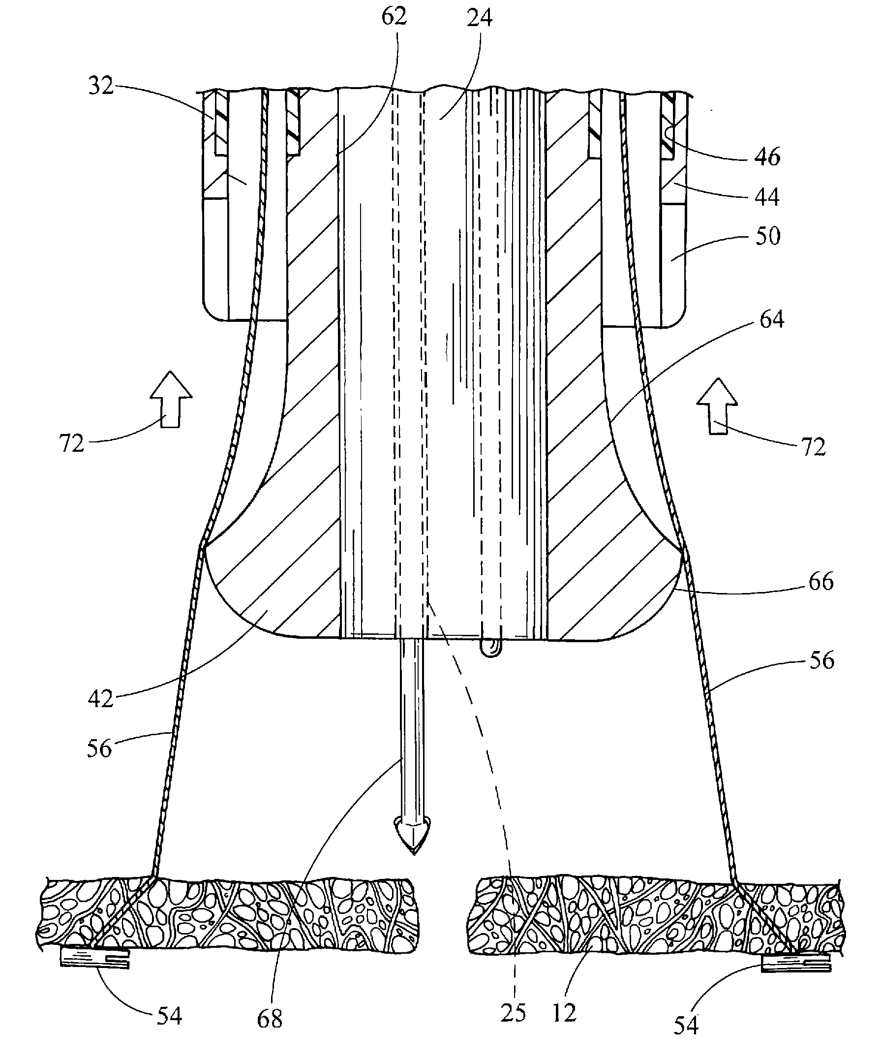
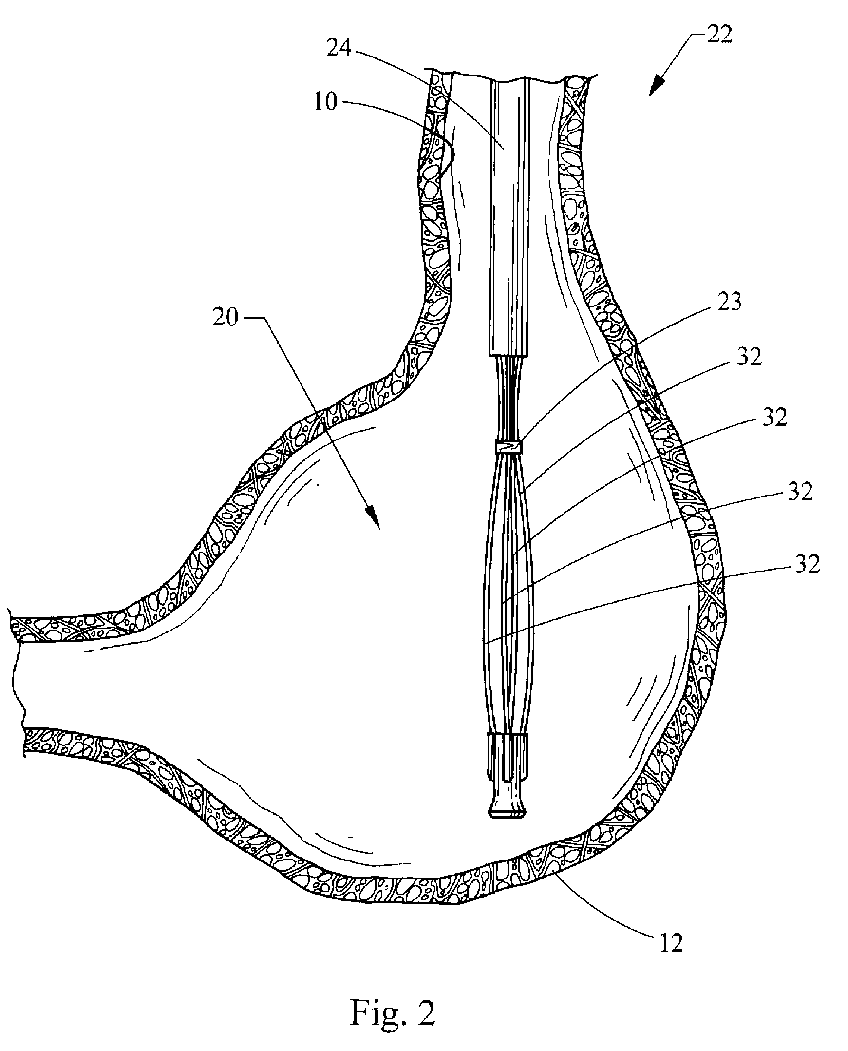
[0016]Turning now to the figures, FIG. 1 depicts a medical device 20 constructed in accordance with the teachings of the present invention. As shown in FIG. 2, the medical device 20 also forms part of a medical system 22, which includes the medical device 20 and an endoscope 24. Generally, the medical device 20 is selectively attachable to the endoscope 24, and the medical system 22 may be traversed through a bodily lumen of a patient to a desired location for performing procedures within the body, such as at a particular bodily wall or tissue. As depicted in FIG. 2, the bodily lumen may be the esophagus 10 while the bodily tissue may be the gastric wall 12, although the medical system 22 may be used with any bodily lumen and tissue, as will be understood by those skilled in the art.

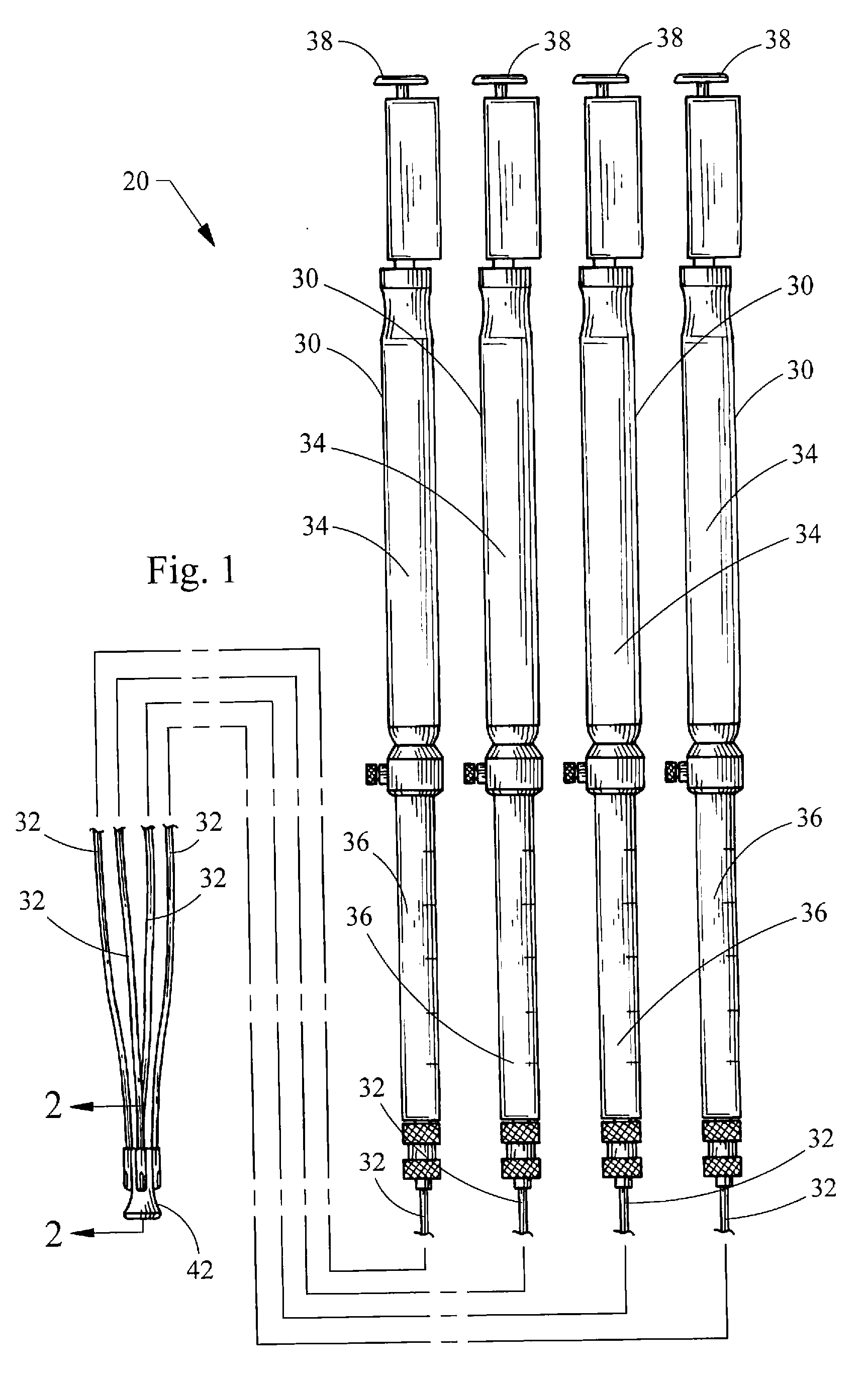
[0017]Turning back to FIG. 1, the medical device 20 generally includes a plurality of puncture needles 30 and a plurality of tubes 32. The plurality of needles 30 have at their proximal end a plurality o...


PUM


Abstract

Medical devices, systems and methods for placing tissue fixation devices are provided. The medical device includes a plurality of elongate delivery devices, a plurality of elongate tubes, and a distal tip. Each delivery device defines a delivery lumen sized to receive a tissue fixation device. Each elongate tube defines a tube lumen sized to receive one of the delivery devices. Each tube lumen has a distal port. The distal tip is connected to the plurality of elongate tubes and defines a guide surface positioned distally of the distal ports. The guide surface is structured to redirect the plurality of delivery devices radially outwardly as the plurality of delivery devices are distally translated through the tube lumens and the distal ports of the plurality of elongate tubes. A medical system is formed by an endoscope that is selectively attachable to the medical device.

Description

CROSS-REFERENCE TO RELATED APPLICATIONS[0001]This application claims the benefit of U.S. Provisional Application Ser. No. 60 / 940,246 filed on May 25, 2007, entitled “MEDICAL DEVICES, SYSTEMS AND METHODS FOR CLOSING PERFORATIONS,” the entire contents of which are incorporated herein by reference.FIELD OF THE INVENTION[0002]The present invention relates generally to medical devices, systems, and methods for closing perforations in tissue.BACKGROUND OF THE INVENTION[0003]Perforations in the walls of internal organs and vessels may be naturally occurring, or formed intentionally or unintentionally. In order to permanently close these perforations and allow the tissue to properly heal, numerous tissue fixation devices and methods have been developed employing sutures, adhesives, clips, staples and the like. One class of such devices is commonly referred to as T-anchors, also known as tissue anchors or visceral anchors. An exemplary tissue anchor is disclosed in U.S. Pat. No. 5,123,914, t...

Claims


Application Information

Patent Timeline
Patent Type & Authority Applications(United States)
IPC IPC(8): A61B17/04A61B1/00
CPCA61B17/0057A61B17/0401A61B17/0469A61B17/0482A61B2017/00637A61B2017/00663A61B2017/0472
Inventor SURTI, VIHAR C.
Owner COOK MEDICAL TECH LLC
Why Eureka
Try Eureka