Edge detection in ultrasound images

a technology of ultrasound images and edge detection, applied in the field of medical imaging, can solve the problems of requiring substantial user involvement and difficult segmentation of ultrasound images in order to find 3-dimensional contours, and achieve the effect of improving edge detection

Inactive Publication Date: 2009-03-26
BIOSENSE WEBSTER INC
View PDF31 Cites 68 Cited by
  • Summary
  • Abstract
  • Description
  • Claims
  • Application Information

AI Technical Summary

Benefits of technology

[0007]Embodiments of the present invention improve edge detection in 2-dimensional image data, e.g., ultrasound image data, that may be carried out automatically with minimal user involvement. The methods of edge detection in accordance with these embodiments are carried out nearly automatically, using an image processing technique that results in generation of a segmented edge contour, which may then be used in 3-dimensional reconstruction and segmentation.

Problems solved by technology

Segmentation of ultrasound images in order to find 3-dimensional contours remains a difficult task, which generally requires substantial user involvement.

Method used


Image

  • Edge detection in ultrasound images
  • Edge detection in ultrasound images
  • Edge detection in ultrasound images

Examples


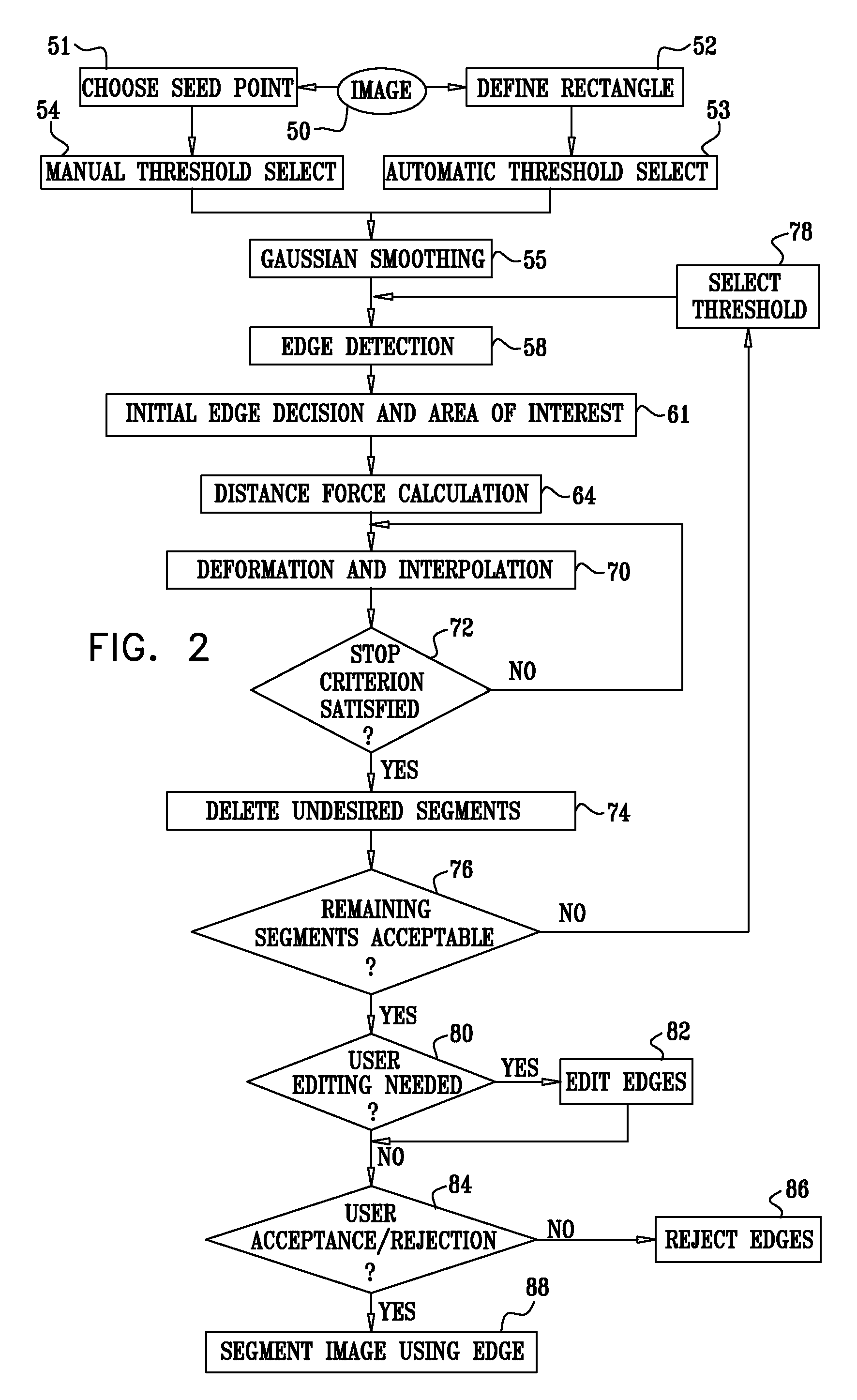
Embodiment Construction

[0026]In the following description, numerous specific details are set forth in order to provide a thorough understanding of the present invention. It will be apparent to one skilled in the art, however, that the present invention may be practiced without these specific details. In other instances, well-known circuits, control logic and the details of computer program instructions for conventional algorithms and processes have not been shown in detail in order not to obscure the present invention unnecessarily.

[0027]Software programming code, which embodies aspects of the present invention, is typically maintained in permanent storage, such as a computer readable medium. In a client / server environment, such software programming code may be stored on a client or a server. The software programming code may be embodied on any of a variety of known tangible media for use with a data processing system, such as a diskette, or hard drive, or CD-ROM. The code may be distributed on such media...


PUM


Abstract

Embodiments of the present invention improve edge detection in 2-dimensional image data that may be carried out automatically with minimal user involvement. The invention is carried automatically, using an image processing technique that results in generation of a segmented edge contour, which may then be used in 3-dimensional reconstruction and segmentation.

Description

CROSS-REFERENCE TO RELATED APPLICATIONS[0001]This application claims the benefit of U.S. Provisional Application No. U.S. 60 / 915,152, filed May 1, 2007, which is herein incorporated by reference.BACKGROUND OF THE INVENTION[0002]1. Field of the Invention[0003]This invention relates to medical imaging. More particularly, this invention relates to improvements in edge detection of intrabody structures in ultrasound images.[0004]2. Description of the Related Art[0005]Ultrasound imaging is now well established as a modality for imaging structures in the body, such as the heart. For example, U.S. Pat. No. 6,066,096, whose disclosure is incorporated herein by reference, describes an imaging probe for volumetric intraluminal ultrasound imaging. The probe, configured to be placed inside a patient's body, includes an elongated body having proximal and distal ends. An ultrasonic transducer phased array is connected to and positioned on the distal end of the elongated body. The ultrasonic trans...

Claims


Application Information

Patent Timeline
Patent Type & Authority Applications(United States)
IPC IPC(8): G06K9/00G06K9/46
CPCG06T7/0085G06T2207/30048G06T2207/10132G06T7/0089A61B8/4254G06T7/13G06T7/149A61B8/00
Inventor ZUR, DRORMAYZLISH, DOVPATT, ZAFRIR
Owner BIOSENSE WEBSTER INC
Why Eureka
Try Eureka