Method of Enhancing Ultrasound Visibility of Hyperechoic Materials

a hyperechoic marker and ultrasound technology, applied in the field of improving the visibility of hyperechoic markers, can solve the problems of inability to use biodegradable markers, inability to use sutures and collagen-based markers, and inability to surround hyperechoic markers with water, so as to achieve easy visualization and better retain markers

Inactive Publication Date: 2009-04-02
DEVICOR MEDICAL PROD
View PDF13 Cites 7 Cited by
  • Summary
  • Abstract
  • Description
  • Claims
  • Application Information

AI Technical Summary

Benefits of technology

[0025]The hypoechoic nature of the hydrated plug enables ultrasound visibility of the permanent, temporary or combination of permanent and temporary markers within the hydrogel hydrated plug because the permanent, temporary, or permanent / temporary combination marker is outlined as a specular reflector within a hypoechoic hydrated marker plug having a water-like nonreflective substrate.
[0026]Water is the most easily visualized substrate under ultrasound. The permanent, temporary, or combination of permanent and temporary markers of this invention can have any shape that are not easily confused with a natural shape as mentioned above and it can be made of any permanent metallic-like or hard plastic material. Helical shapes having a hollow interior is a preferred shape because it allows the polymer to better retain the marker within.

Problems solved by technology

Biodegradable markers are not permanent and therefore cannot be relied upon if a biopsy site is to be re-located at a time remote from the time of the biopsy.
Suture and collagen-based markers are not suitable as markers because they are hyperechoic, i.e., difficult to see under ultrasound because such materials are easily confused with other shadowing normal structures in the body such as fibrous tissue, fatty tissue, ducts in breast tissue, and the like, for example.
However, the art includes no means for surrounding a hyperechoic marker with water at a biopsy site.

Method used


Image

  • Method of Enhancing Ultrasound Visibility of Hyperechoic Materials
  • Method of Enhancing Ultrasound Visibility of Hyperechoic Materials
  • Method of Enhancing Ultrasound Visibility of Hyperechoic Materials

Examples


Embodiment Construction

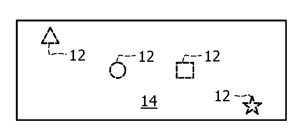
[0045]Referring now to FIGS. 1A and 1B, it will there be seen that a first illustrative embodiment of the invention is denoted as a whole by the reference numeral 10.

[0046]Hydrogel plug 10 includes a plurality of temporary markers, collectively denoted 12, embedded within a hydrogel material 14 having, in this first embodiment, a shape designed to inhibit migration of the plug within tissue. The FIG. 1A configuration is the “in repose” configuration of plug 10. Markers 12 may be formed of metal, hard plastic, or other permanent material but the depicted markers 12 are formed of a temporary, biodegradable materials.

[0047]FIGS. 1A and 1B depict multiple temporary markers 12 of differing sizes and shapes just to illustrate that the markers may be provided in an infinite variety of geometrical configurations. Thus it should be understood that a single temporary marker 12 is within the scope of this invention. Accordingly, the novel method of this invention includes the step of selecting...


PUM

PropertyMeasurementUnit
sizeaaaaaaaaaa
shapeaaaaaaaaaa
biocompatibleaaaaaaaaaa

Abstract

A permanent or temporary marker is embedded within a hydrogel plug used in a biopsy procedure to indicate the location of a suspicious lesion so that the marker may be found in a subsequent surgical procedure. A combination of at least one permanent and at least one temporary marker may be used. Inserts of differing sizes and shapes may also be placed into the hydrogel plugs during their manufacturing process and removed from the plugs after the plugs have cured so that air-filled cavities are left in the plug. These cavities are temporary but may be used to augment location of a permanent marker.

Description

CROSS-REFERENCE TO RELATED DISCLOSURES[0001]This disclosure is a continuation-in-part of U.S. patent application Ser. No. 11 / 277,721, filed Mar. 28, 2006, by the same inventors and having the same title. That disclosure is hereby incorporated by reference in its entirety into this disclosure.BACKGROUND OF THE INVENTION[0002]1. Field of the Invention[0003]This invention relates, generally, to a method for improving the visibility of a hyperechoic marker or markers under ultrasound. Such markers are used to indicate the location of a tumor or lesion so that a procedure to remove such lesion or tumor may be performed weeks or months after the marker has been implanted. More particularly, it relates to markers that incorporate hydrogels to enhance the visibility of the markers with imaging techniques such as ultrasound and to methods for making such markers.[0004]2. Description of the Prior Art[0005]A permanent metal or hard plastic, such as a permanent, bio-compatible plastic such as p...

Claims


Application Information

Patent Timeline
Patent Type & Authority Applications(United States)
IPC IPC(8): A61B8/00
CPCA61B19/54A61B2019/5425A61B2017/00004A61B2017/00526A61B2017/00898A61B90/39A61B2090/3925B29C39/10
Inventor FISHER, JOHN S.
Owner DEVICOR MEDICAL PROD
Why Eureka
Try Eureka