
3D General Lesion Segmentation In CT

Active Publication Date: 2009-04-16
SIEMENS HEALTHCARE GMBH
View PDF41 Cites 31 Cited by
  • Summary
  • Abstract
  • Description
  • Claims
  • Application Information

AI Technical Summary

Benefits of technology

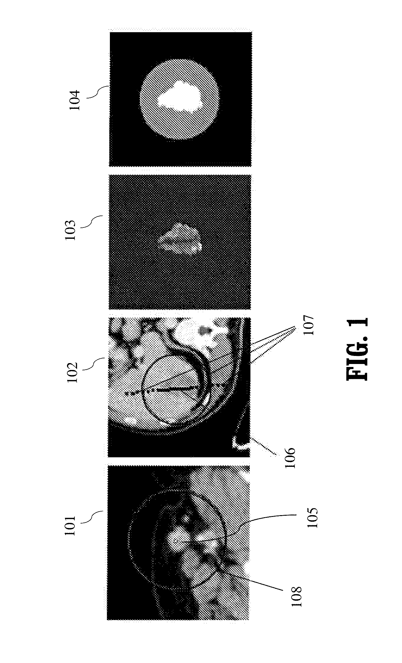
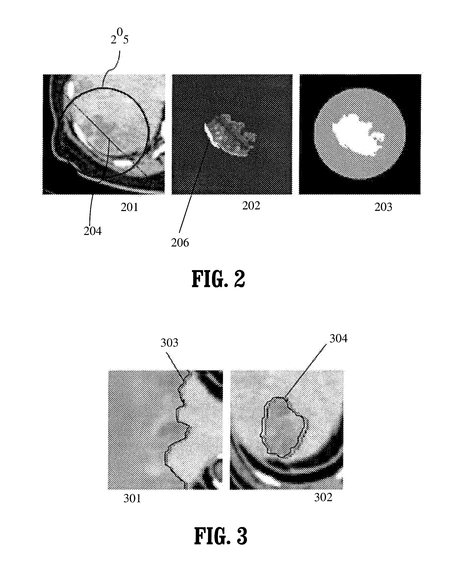
[0019]In accordance with another aspect of the present invention, a method is provided for segmenting an object from a background in three-dimensional (3D) image data, wherein determ

Problems solved by technology

Unfortunately, there are no good tools available to measure lesion volumes and it is very time consuming to manually outline them in 3D.

Method used


Image

  • 3D General Lesion Segmentation In CT
  • 3D General Lesion Segmentation In CT
  • 3D General Lesion Segmentation In CT

Examples


Embodiment Construction

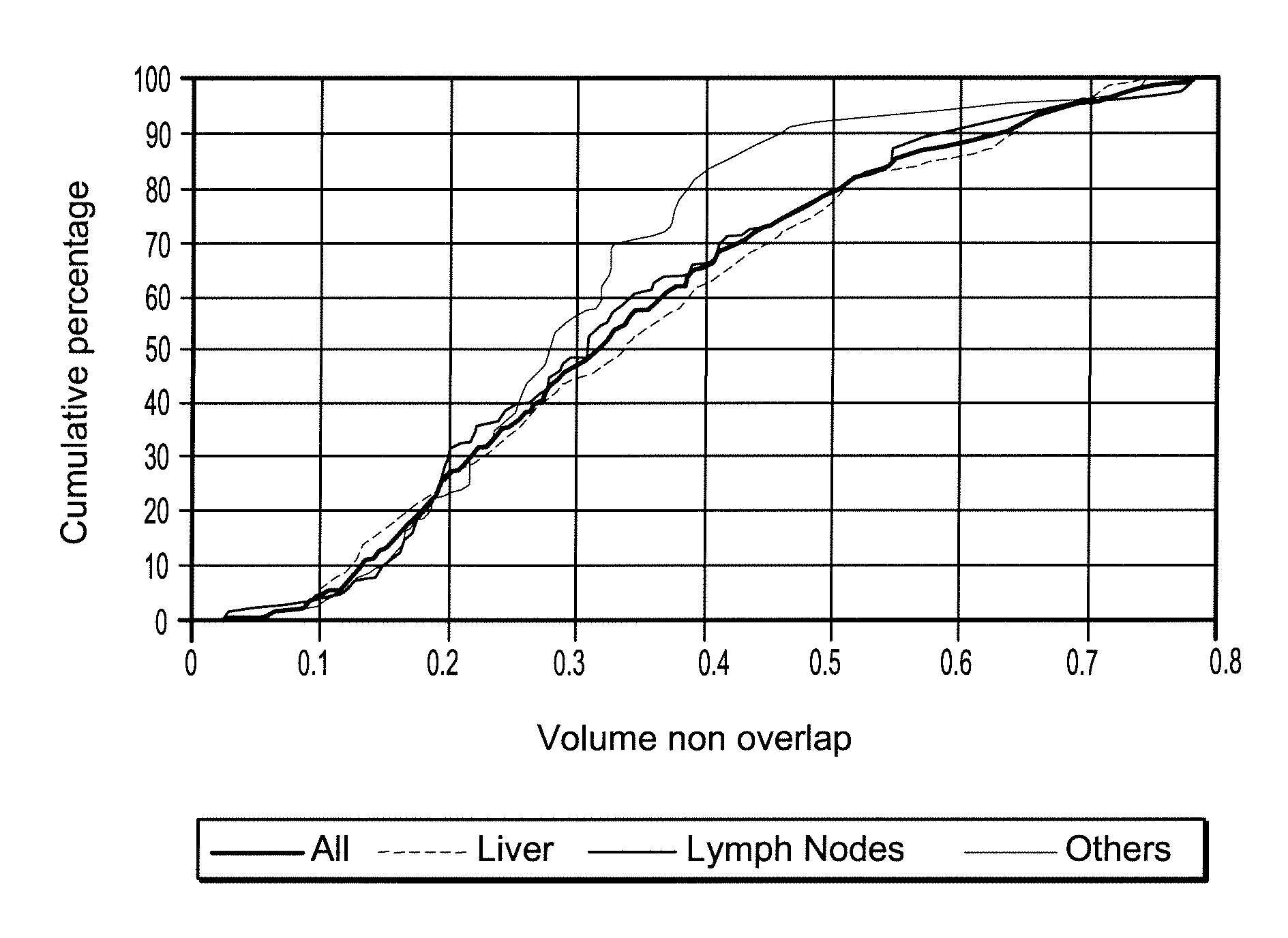
[0037]Most of the work on medical analysis for cancer screening and treatment has been in the context of lung cancer, mammography, and colon cancer. There has been very little interest in liver lesion segmentation though some research is described in P. J. Yim and D. J. Foran, “Volumetry of hepatic metastases in computed tomography using the watershed and active contour algorithms,” in Proc. IEEE Symposium on Computer-Based Medical Systems, New York, N.Y., 2003, pp. 329-335, and in M. Bilello, S. B. Gokturk, T. Desser, S. Napel, R. B. Jeffrey Jr., and C. F. Beaulieu, “Automatic detection and classification of hypodense hepatic lesions on contrast-enhanced venous-phase CT,” Medical Physics, vol. 31, no. 9, pp. 2584-2593, 2004. Earlier work has focused on relatively simple hypodense liver tumors or metastases which are nicely contrasted against the parenchyma. Papers have presented very simple image processing techniques (thresholding, watershed, active contours) which have been teste...


PUM


Abstract

A general purpose method to segment any kind of lesions in 3D images is provided. Based on a click or a stroke inside the lesion from the user, a distribution of intensity level properties is learned. The random walker segmentation method combines multiple 2D segmentation results to produce the final 3D segmentation of the lesion.

Description

CROSS-REFERENCE TO RELATED APPLICATIONS[0001]This application claims the benefit of U.S. Provisional Application No. 60 / 978,818, filed Oct. 10, 2007, which is incorporated herein by reference.BACKGROUND OF THE INVENTION[0002]The present invention relates to methods and apparatus for image segmentation for separation of a foreground object from a background. In particular, it relates to interactive 3D segmentation of an object from a background.[0003]Cancer is the second leading cause of death in the United States as half a million people die each year. For this reason, it is important to diagnose the disease as early as possible and monitor it carefully as it is being treated. In addition to primary tumors, physicians are also interested in secondary tumors that might have metastasized through the rest of the body. The main areas of concern are the liver (because many forms of cancer generate liver metastases) and lymph nodes (because cancers spread through the lymphatic system).[00...

Claims


Application Information

Patent Timeline
IPC IPC(8): G06K9/80
CPCA61B6/466G06T7/0083G06T7/0087G06T2207/10081G06T2207/30096G06T2207/20096G06T2207/20101G06T2207/20136G06T2207/20076G06T7/12G06T7/143G06T7/181
Inventor JOLLY, MARIE-PIERREGRADY, LEO
Owner SIEMENS HEALTHCARE GMBH
Learn More