Method for Preparing Cell Fraction Containing Hemangioblasts

a cell fraction and hemangioblast technology, applied in the field of hemangioblasts, can solve the problems of unexplored nature of hemangioblasts, defects in both hematopoiesis and vasculogenesis, and still remains unknown how ltr-hscs in the yolk sac acquire full repopulation activity, so as to facilitate purification and detection of proteins

Inactive Publication Date: 2009-05-21
TOUDAITLO LTD
View PDF2 Cites 1 Cited by
  • Summary
  • Abstract
  • Description
  • Claims
  • Application Information

AI Technical Summary

Benefits of technology

[0070]The protein of this invention can be prepared as either a natural protein or a recombinant protein utilizing gene recombination techniques. Natural proteins can be prepared by, for example, subjecting extracts from tissues and cells that are presumed to express mouse PCLP1 protein (for example, LO cells, embryo cells in AGM region, etc.) to affinity chromatography using the above-described antibody to the mouse PCLP1 protein. On the other hand, recombinant proteins may be prepared by culturing cells transformed with DNA encoding the mouse PCLP1 protein, allowing the transformants to express the protein, and recovering the protein as described below. Proteins of this invention can be fused to peptide tags and other proteins. Such fusion proteins can be useful for facilitating the purification and detection of proteins.

Problems solved by technology

However, LTR-HSCs, which are capable of repopulating lethally irradiated adult mice, have not been found in the P-Sp region.
However, it still remains unknown how LTR-HSCs in the yolk sac acquire full repopulation activity.
Furthermore, gene disruption of the SCL / Tal-1 transcription factor caused defects in both hematopoiesis and vasculogenesis, which were similar to those of Flk1 knockout mice (Porcher, C. et al.
However, the nature of hemangioblasts remains unexplored, in particular, there is yet no absolute evidence that LTR-HSCs are derived from hemangioblasts.

Method used


Image

  • Method for Preparing Cell Fraction Containing Hemangioblasts
  • Method for Preparing Cell Fraction Containing Hemangioblasts
  • Method for Preparing Cell Fraction Containing Hemangioblasts

Examples


example 2

Preparation of Monoclonal Antibodies Against Surface Antigens of Endothelial-Like Cells Derived from AGM Culture

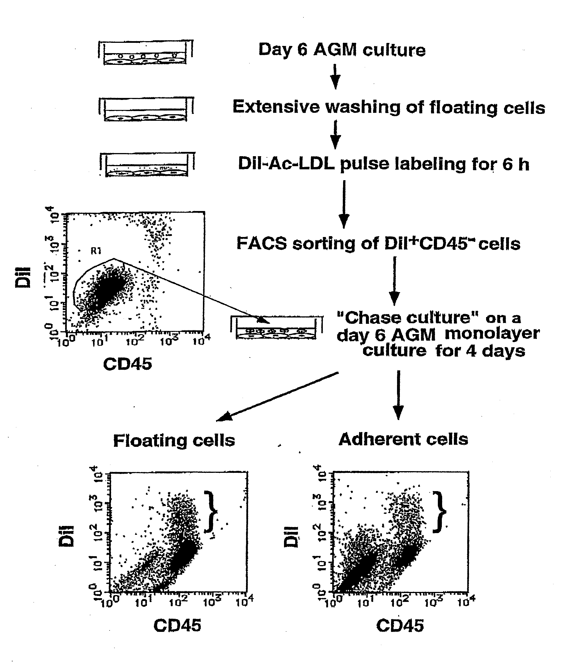
[0119]To define hemangioblasts more precisely, the inventors aimed to obtain a specific antibody directed against hemangioblasts. By repeating the passage of adherent cells of the AGM culture in the presence of OSM, the inventors were able to establish a novel OSM-dependent endothelial-like cell line, LO. LO cells exhibit characteristics very similar to those of endothelial-like cells in the AGM culture, such as endothelial-like morphology, incorporation of DiI-Ac-LDL, and production of hematopoietic cells. The inventors used the LO cells as immunogens to raise monoclonal antibodies against cell surface antigens on LO cells as follows.

[0120]Wistar rats (Nihon SLC) were immunized with 107 of LO cells in the presence of Freund's complete adjuvant (WAKO, Osaka, Japan) according to the standard immunization procedure (Hockfield, S. et al. (1993) “Selected Methods for Antibody ...

example 3

Molecular Cloning of Mouse PCLP1 Molecule as a Possible Hemangioblast Antigen

[0122]Next, using a standard expression cloning strategy with COS7 cells and 10B9 monoclonal antibody, the inventors isolated a cDNA clone encoding the 10B9 antigen.

[0123]Expression cloning of a cDNA encoding the 10B9 antigen was carried out by using COS7 cells as previously described (Harada, N. et al. (1990) Proc. Natl. Acad. Sci. USA 87, 857-861) except that magnetic beads conjugated with anti-rat IgG antibody (Dynabeads M-450) (Dynal, Oslo, Norway) were employed instead of plate panning. Briefly, COS7 cells were fused with spheroplasts of the cDNA plasmid library of LO cells (Tanaka, M. et al. (1999) Blood 93, 804-815) and stained with 10B9 antibody followed by Dynabead selection. Plasmid DNA mixture was harvested from the beads, amplified in E. coli and re-transfected into COS7 cells. This procedure was repeated 4 to 5 times until a single band of cDNA insert was recovered. As a result, the inventors i...

example 4

Expression of PCLP1 on the Endothelial-Like Cells in the AGM Culture

[0128]The inventors examined the expression of PCLP1 on the endothelial-like cells in the AGM culture by immunostaining with 10B9 anti-PCLP1 antibody. Cultured AGM-derived cells in plastic plates were fixed with 1% paraformaldehyde (PFA)-PBS at room temperature for 15 minutes and incubated with anti-PCLP1 10B9 antibody at 10 μg / ml at 4° C. over night. After incubation with peroxidase-conjugated anti-rat IgG (Amersham), signals were visualized by 3,3′-diaminobenzidine (DAB) as previously described (Hara, T. et al. (1998) Dev. Biol. 201, 144-153).

[0129]As the inventors expected, PCLP1 was detectable on endothelial-like cells (FIG. 5A to C), but not on fibroblastic cells (data not shown) in the AGM culture. The endothelial-like cells, defined by their polygonal cell morphology and incorporation of DiI-Ac-LDL, were further fractionated by fluorescent activated cell sorting (FACS) using anti-PCLP1 and anti-CD45 antibodie...


PUM

PropertyMeasurementUnit
pHaaaaaaaaaa
densityaaaaaaaaaa
temperatureaaaaaaaaaa

Abstract

Mouse PCLP1 was identified by expression cloning with the use of a monoclonal antibody against a surface antigen of a cell line derived from mouse AGM. By fractionating PCLP1-positive / CD45-negative cells and culturing them in vitro, it was clarified that these cells differentiate into endothelial-like cells, angioblast-like cells, and hematopoietic cells. By transferring the PCLP1-positive / CD45-negative cells into a mouse defective in the hematopoietic function, the hematopoietic system was reconstructed over a long period of time. These facts indicate that the PCLP1-positive / CD45-negative cells contain mammalian hemangioblasts capable of expressing the activity as long-term repopulating hematopoietic stem cells (LTR-HSC). The present invention provides a method for preparing a cell fraction containing hemangioblasts, the cell fraction prepared by the method, and use of this cell fraction.

Description

RELATED APPLICATIONS[0001]This application is a divisional application of U.S. application Ser. No. 10 / 130,076, filed May 10, 2002, which claims benefit of priority to Japan Patent Application No. 11-320234, filed Nov. 10, 1999, and PCT International Application No. PCT / JP00 / 07817, filed Nov. 7, 2000.TECHNICAL FIELD[0002]The present invention relates to a marker molecule for hemangioblasts, method for preparing a cell fraction containing hemangioblasts using the marker molecule, cell fraction prepared by the method, and use of the cell fraction.BACKGROUND ART[0003]Development of hematopoiesis proceeds through two distinct steps, i.e. primitive and definitive hematopoiesis. In mice, primitive hematopoiesis begins in the extraembryonic yolk sac at 7.5 days post coitum (dpc) in gestation, while definitive hematopoiesis, which is distinguished by enucleated erythrocytes, lymphopoiesis, and generation of long term repopulating hematopoietic stem cells (LTR-HSCs), originates from the intr...

Claims


Application Information

Patent Timeline
Patent Type & Authority Applications(United States)
IPC IPC(8): C07K14/47C07K7/06C07K7/08A01K67/027C07K16/18C07K16/28C12N5/074C12N5/0789C12N15/12C12P21/08
CPCA01K67/0271C07K16/18C07K16/28C12N5/0647C12N5/0692C12N2501/115C12N2502/1394C12N2501/14C12N2501/165C12N2501/2303C12N2501/235C12N2501/237C12N2501/125
Inventor MIYAJIMA, ATSUSHIHARA, TAKAHIKO
Owner TOUDAITLO LTD
Why Eureka
Try Eureka