Generating MRI images usable for the creation of 3D bone models employed to make customized arthroplasty jigs

a technology of mri and mri scan, which is applied in the field of medical imaging, can solve the problems of increasing the radiation dose to a patient, adverse reactions in some patients, and ct imaging not providing the image resolution of mri

Active Publication Date: 2009-05-28
HOWMEDICA OSTEONICS CORP
View PDF114 Cites 387 Cited by
  • Summary
  • Abstract
  • Description
  • Claims
  • Application Information

AI Technical Summary

Problems solved by technology

Creating the 2D images or image slices via CT imaging is relatively quick, but involves multiple scans, thus increasing the dose of radiation to a patient.
Also, CT imaging requires the use of a contrast agent, which may cause an adverse reaction in some patients.
Finally, CT imaging does not provide the image resolution offered by MRI.
Unfortunately, conventional MRI is not quick, requiring a patient to maintain a position completely still for 30, 45 or more minutes in order to obtain image slices that have adequate resolution.
It is often extremely difficult, if not impossible, for a patient to rema...

Method used


Image

  • Generating MRI images usable for the creation of 3D bone models employed to make customized arthroplasty jigs
  • Generating MRI images usable for the creation of 3D bone models employed to make customized arthroplasty jigs
  • Generating MRI images usable for the creation of 3D bone models employed to make customized arthroplasty jigs

Examples


Embodiment Construction

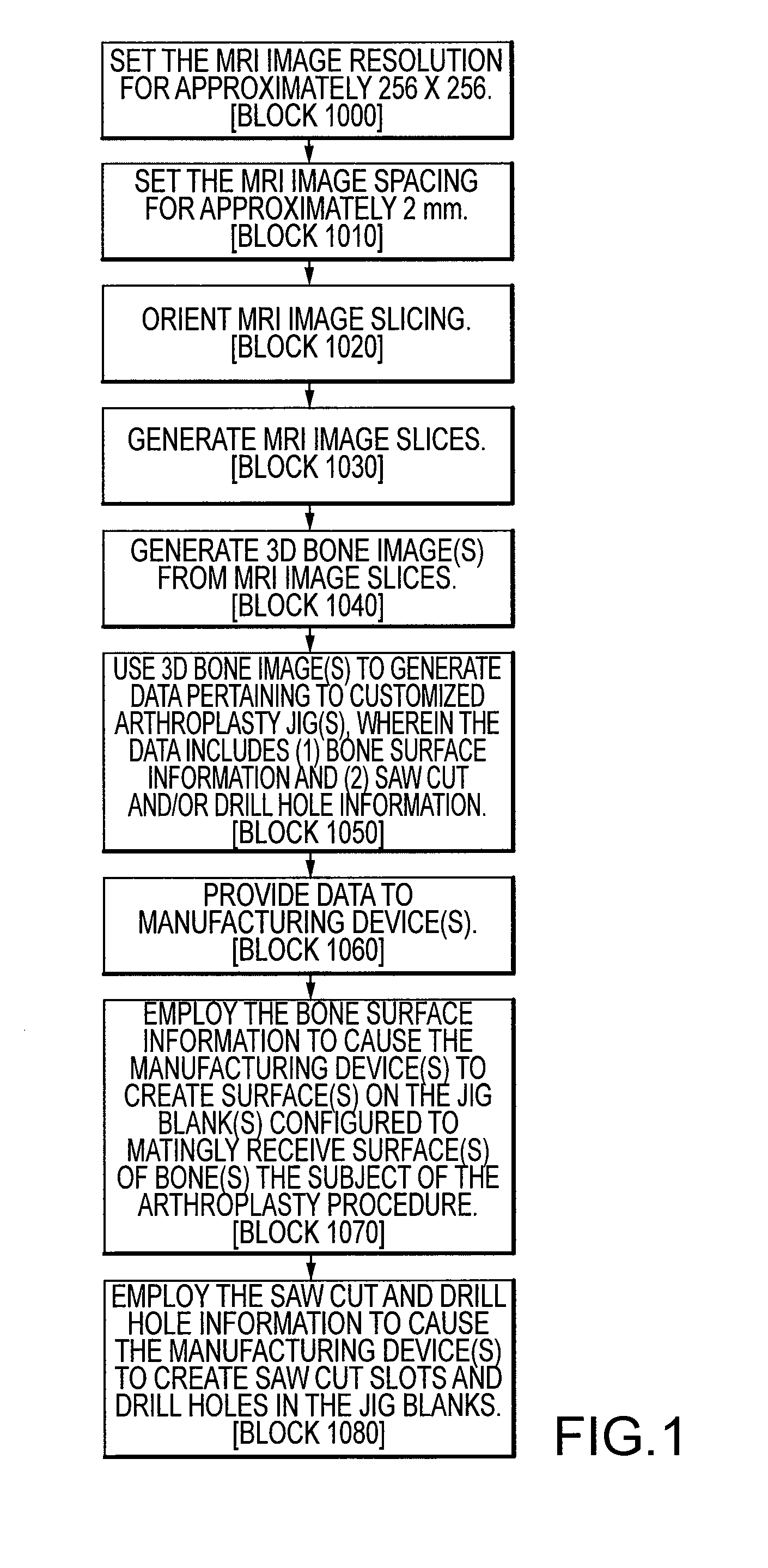
[0032]Disclosed herein is a system and method for generating 2D MRI image slices 10 that are readily and reliably useable in creating 3D bone models 15 used in the generation of customized arthroplasty jigs 20. A MRI image resolution and MRI image slice spacing SS disclosed herein significantly reduce the time required to generate a series of MRI images slices 10, thereby making it possible for a patient 22 to maintain a position completely still for the duration of the MRI imaging process. An image slice orientation method disclosed herein ensures the 2D MRI image slices 10 are readily and reliably useable with a 3D computer modeling program to create 3D bone models 15 used in the generation of customized arthroplasty jigs 20.

[0033]For a discussion of the overall process of turning MRI image slices 10 into a customized arthroplasty jig 20, reference is made to FIGS. 1 and 2. FIG. 1 is a flow chart illustrating a method of creating customized arthroplasty jigs 20 for mating with sur...


PUM


Abstract

Disclosed herein is a method of creating a customized arthroplasty jig. The method may include: generating two-dimensional MRI images of a patient's joint area to undergo arthroplasty; electronically orienting the two dimensional MRI image slices to account for the patient's joint area being randomly physically oriented in a scanning area of a MRI machine; generating a three-dimensional bone image of at least a portion of a bone of the patient's joint area from the generated two-dimensional MRI images; using the three-dimensional bone image to generate data pertaining to the customized arthroplasty jig, wherein the data includes bone surface information; providing the data to at least one manufacturing device; and employing the bone surface information to cause the at least one manufacturing device to create a surface on the arthroplasty jig configured to matingly receive a surface of the bone.

Description

FIELD OF THE INVENTION[0001]The present invention relates to medical imaging. More specifically, the present invention relates to medical imaging employed to generate three-dimensional bone models for use in the creation of customized arthroplasty jigs.BACKGROUND OF THE INVENTION[0002]Systems and methods for making customized arthroplasty jigs are disclosed in these three U.S. patent applications: U.S. patent application Ser. No. 11 / 656,323 to Park et al., titled “Arthroplasty Devices and Related Methods” and filed Jan. 19, 2007; U.S. patent application Ser. No. 10 / 146,862 to Park et al., titled “Improved Total Joint Arthroplasty System” and filed May 15, 2002; and U.S. patent Ser. No. 11 / 642,385 to Park et al., titled “Arthroplasty Devices and Related Methods” and filed Dec. 19, 2006. The disclosures of these three U.S. patent applications are incorporated by reference in their entireties into the present patent application.[0003]As explained in U.S. patent application Ser. No. 11 / ...

Claims


Application Information

Patent Timeline
IPC IPC(8): A61B17/58A61B5/055
CPCA61B5/055A61B5/4528A61B17/154A61B2019/5236A61B17/157A61B19/50A61B2019/505A61B17/155A61B34/10A61B2034/105A61B2090/374
Inventor PARK, ILWHANHOWELL, STEPHEN M.
Owner HOWMEDICA OSTEONICS CORP
Why Eureka
Try Eureka