Magnetic resonance imaging apparatus and magnetic resonance imaging method

a magnetic resonance imaging and magnetic resonance imaging technology, applied in the direction of material analysis using wave/particle radiation, instruments, nuclear engineering, etc., can solve the problems of image degradation, image wobble, and displaced slice position, and achieve high resolution

Active Publication Date: 2009-06-11
TOSHIBA MEDICAL SYST CORP
View PDF12 Cites 15 Cited by
  • Summary
  • Abstract
  • Description
  • Claims
  • Application Information

AI Technical Summary

Benefits of technology

[0015]The magnetic resonance imaging apparatus and the magnetic resonance imaging method according to the present invention as des

Problems solved by technology

However, in the conventional cardiac cine imaging with MRI, there are problems that image degradation such as image wobble occurs and a slice position is displaced in the case of imaging an image of a patient that is difficult to hold his breath.
As a result, there is concern that accuracy of diagnostic such as cardiac function analysis decreases.
In the conventional cardiac cine

Method used


Image

  • Magnetic resonance imaging apparatus and magnetic resonance imaging method
  • Magnetic resonance imaging apparatus and magnetic resonance imaging method
  • Magnetic resonance imaging apparatus and magnetic resonance imaging method

Examples


Embodiment Construction

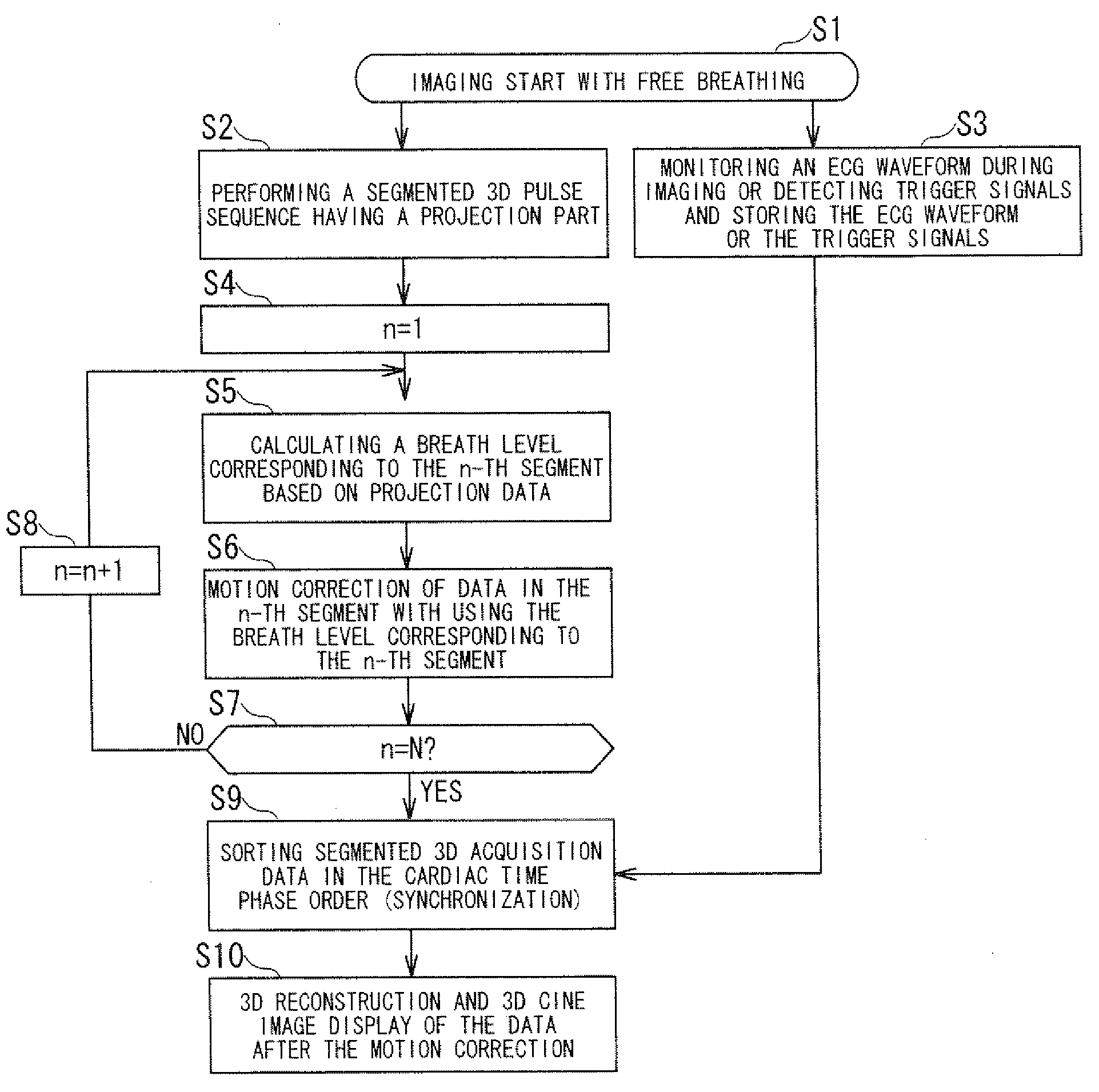
[0024]A magnetic resonance imaging apparatus and a magnetic resonance imaging method according to embodiments of the present invention will be described with reference to the accompanying drawings.

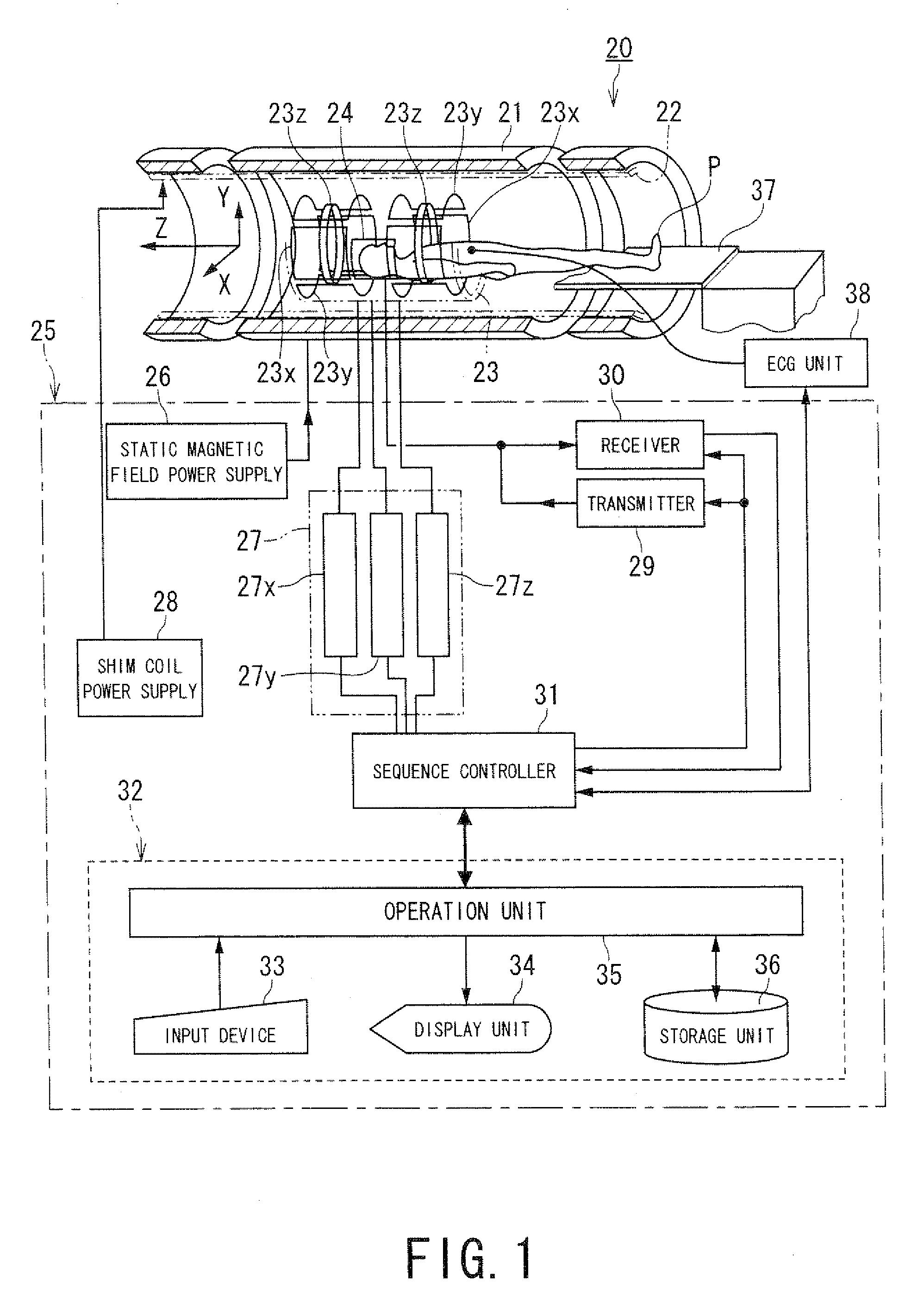
[0025]FIG. 1 is a block diagram showing a magnetic resonance imaging apparatus according to an embodiment of the present invention.

[0026]A magnetic resonance imaging apparatus 20 includes a static field magnet 21 for generating a static magnetic field, a shim coil 22 arranged inside the static field magnet 21 which is cylinder-shaped, a gradient coil 23 and a RF coil 24. The static field magnet 21, the shim coil 22, the gradient coil 23 and the RF coil 24 are built in a gantry (not shown).

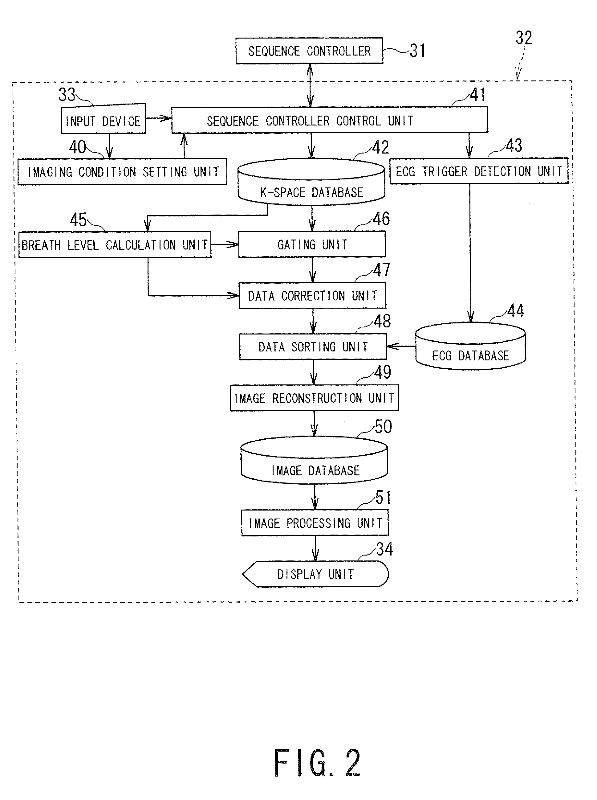
[0027]The magnetic resonance imaging apparatus 20 also includes a control system 25. The control system 25 includes a static magnetic field power supply 26, a gradient power supply 27, a shim coil power supply 28, a transmitter 29, a receiver 30, a sequence controller 31 and a computer 32. The gradient po...


PUM


Abstract

A magnetic resonance imaging apparatus includes a data acquisition unit, a correction unit, a sorting unit and n image reconstruction unit. The data acquires data for imaging and projection data. The correction unit performs motion correction of the data using the breath levels obtained based on the projection data. The data sorting unit sorts the data after the motion correction into a cardiac time phase order based on electrocardiographic information. The image reconstruction unit reconstructs three dimensional image data based on the sorted data after the motion correction.

Description

BACKGROUND OF THE INVENTION[0001]1. Field of the Invention[0002]The present invention relates to a MRI (magnetic resonance imaging) apparatus and a magnetic resonance imaging method which excite nuclear spin of an object magnetically with a RF (radio frequency) signal having the Larmor frequency and reconstruct an image based on NMR (nuclear magnetic resonance) signals generated due to the excitation, and more particularly, to a magnetic resonance imaging apparatus and a magnetic resonance imaging method which make it possible to perform 3D (three-dimensional) cine imaging of a moving part such as a heart under free breath.[0003]2. Description of the Related Art[0004]Magnetic Resonance Imaging is an imaging method which excites nuclear spin of an object set in a static magnetic field with a RF signal having the Larmor frequency magnetically and reconstruct an image based on a MR signal generated due to the excitation.[0005]Conventionally, in the field of MRI, a cardiac cine imaging ...

Claims


Application Information

Patent Timeline
IPC IPC(8): G06T7/00G06T15/00
CPCG01R33/56316G01R33/56308
Inventor YUI, MASAO
Owner TOSHIBA MEDICAL SYST CORP
Why Eureka
Try Eureka