
Bioabsorbable stent with radiopaque layer and method of fabrication

a bioabsorbable polymer and radiopaque layer technology, applied in the field of implantable medical devices, can solve the problems of insufficient radiographic density of commercially available bioabsorbable polymers, and inability to be easily imaged by fluoroscopy

Inactive Publication Date: 2009-06-11
ABBOTT CARDIOVASCULAR
View PDF9 Cites 25 Cited by
  • Summary
  • Abstract
  • Description
  • Claims
  • Application Information

AI Technical Summary

Benefits of technology

"The present invention provides a bioabsorbable stent with a scaffolding that includes a support layer and a radiopaque layer. The stent is made by co-extruding a tube with a radiopaque layer and a support layer, or by forming a radiopaque coating layer over at least a portion of a surface of a tube. The stent can be easily fabricated and has improved visibility during placement. The technical effects of the invention include improved placement accuracy and reduced complications during stent insertion."

Problems solved by technology

However, all commercially available bioabsorbable polymers are radiolucent and do not possess sufficient radiographic density to be easily imaged by fluoroscopy.

Method used


Image

  • Bioabsorbable stent with radiopaque layer and method of fabrication
  • Bioabsorbable stent with radiopaque layer and method of fabrication
  • Bioabsorbable stent with radiopaque layer and method of fabrication

Examples


example 1

[0082]1. A blend of PLLA-co-PCL (90% PLLA and 10% PCL) and tungsten nanoparticles (polymer / particle −20:1 by volume or 1:1 by weight) is prepared using a compounder at 200° C.

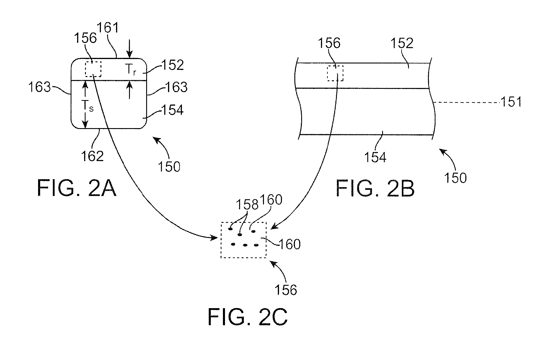
[0083]2. A bi-layer tube is formed by the co-extrusion of PLLA as an inner layer and the PLLA-co-PCL / tungsten nanoparticles as the radiopaque outer layer. The thickness of the PLLA outer layer and the radiopaque layer is set to 0.05 inch and 0.02 inch, respectively. The inside diameter (ID) of the extruded tubing is set to about 0.021 inch and the outside diameter (OD) is about 0.091 inch.

[0084]3. The bi-layer tube is radially expanded. A stent pattern is cut into the expanded tubing. The total thickness of the stent struts is about 0.008 inch.

example 2

[0085]1. A blend of PLLA and barium sulfate (100:30 by weight) is prepared using a compounder at 200° C.

[0086]2. A bi-layer tube is formed by the co-extrusion of PLLA as an inner layer and the PLLA / barium sulfate as the radiopaque outer layer. The thickness of the PLLA outer layer and the radiopaque layer is set to 0.05 inch and 0.02 inch, respectively. The inside diameter (ID) of the extruded tubing is set to about 0.021 inch and the outside diameter (OD) is about 0.091 inch.

[0087]3. The bi-layer tube is radially expanded. A stent pattern is cut into the expanded tubing. The total thickness of the stent struts is about 0.008 inch.

example 3

[0088]1. A blend of PLGA and barium sulfate (100:30 by weight) is prepared using a compounder at 200° C.

[0089]2. A bi-layer tube is formed by the co-extrusion of PLGA as an inner layer and the PLGA / barium sulfate as the radiopaque outer layer. The thickness of the PLGA outer layer and the radiopaque layer is set to 0.05 inch and 0.02 inch, respectively. The inside diameter (ID) of the extruded tubing is set to about 0.021 inch and the outside diameter (OD) is about 0.091 inch.

[0090]3. The bi-layer tube is radially expanded. A stent pattern is cut into the expanded tubing. The total thickness of the stent struts is about 0.008 inch.


PUM


Abstract

Embodiments of a stent and methods of fabricating the same with a bioabsorbable radiopaque layer are disclosed.

Description

BACKGROUND OF THE INVENTION[0001]1. Field of the Invention[0002]This invention relates to implantable medical devices, such as stents. In particular, the invention relates to bioabsorbable polymeric stents with a radiopaque layer.[0003]2. Description of the State of the Art[0004]This invention relates to radially expandable endoprostheses, which are adapted to be implanted in a bodily lumen. An “endoprosthesis” corresponds to an artificial device that is placed inside the body. A “lumen” refers to a cavity of a tubular organ such as a blood vessel. A stent is an example of such an endoprosthesis. Stents are generally cylindrically shaped devices, which function to hold open and sometimes expand a segment of a blood vessel or other anatomical lumen such as urinary tracts and bile ducts. Stents are often used in the treatment of atherosclerotic stenosis in blood vessels. “Stenosis” refers to a narrowing or constriction of the diameter of a bodily passage or orifice. In such treatments...

Claims


Application Information

Patent Timeline
Patent Type & Authority Applications(United States)
IPC IPC(8): A63F9/24
CPCA61L31/12A61L31/18A61L31/148A61B90/39A61B2090/3966A61F2/915
Inventor WANG, YUNBINGGALE, DAVID C.
Owner ABBOTT CARDIOVASCULAR
Learn More