
Methods and Surgical Kits for Minimally-Invasive Facet Joint Fusion

a facet joint and minimally-invasive technology, applied in the field of methods and surgical kits, can solve the problem of out-patient procedur

Inactive Publication Date: 2009-09-17
MINSURG INT
View PDF69 Cites 79 Cited by
  • Summary
  • Abstract
  • Description
  • Claims
  • Application Information

AI Technical Summary

Benefits of technology

[0009]The present invention provides a minimally-invasive procedure and associated surgical tools that can be used to fuse facet joint bones without the use of a screw-based approach. Instead of relying on a screw to hold rubbing facet joints together, the methods of the present invention displaces bone from the facet joint (typically by drilling) and inserts a bone plug into the created hole. The bone plug allows natural bone in-growth into and around the plug such that, typically, a strong and permanent fusion results. The methods of the present invention may be so minimally-invasive that, in some embodiments, it can be practiced arthroscopically or percutaneously. Moreover, in many instances, the procedure can be out-patient.
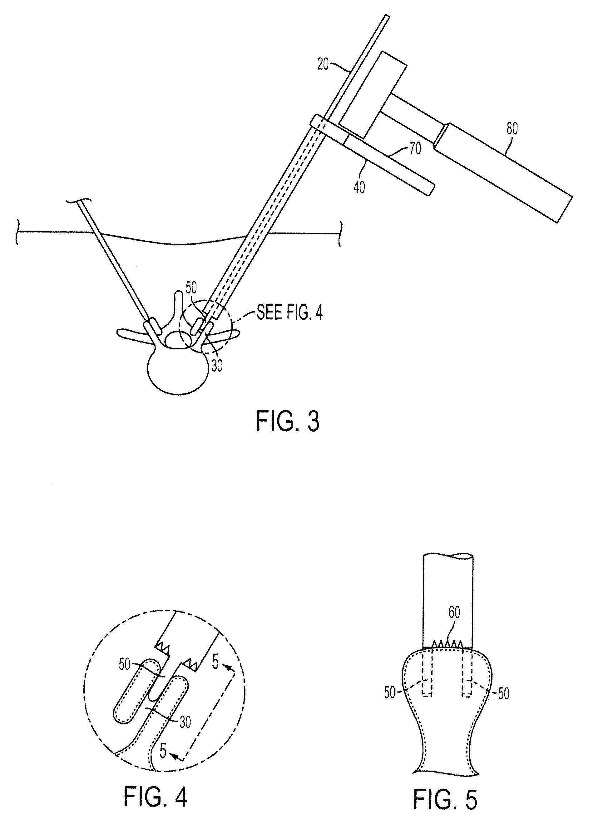
[0028]In some embodiments, the present invention includes a method including: creating an incision; locating a facet joint with a distal end of a pin wherein the facet joint is formed between two opposing bones and the pin comprises the distal end and a proximal end; sliding a substantially hollow drill guide over the pin wherein the drill guide comprises a proximal end, a distal end, and a handle wherein the handle is nearer to the proximal end of the drill guide than to the distal end, the distal end comprises opposed teeth with a length longer than about 3 mm that can be inserted into the facet joint; removing the pin from within the drill guide; inserting a drill bit into the drill guide; drilling a hole into a bone of the facet joint; removing the drill bit; inserting a facet joint bone plug into the hole using a bone plug inserter having a raised portion at or near is proximal end, wherein the raised portion prevents over-insertion of the bone plug; and removing the drill guide. The opposed teeth may be about 3 mm to about 20 mm in length. In one example, the opposed teeth are about 10 mm in length.
[0033]In various embodiments, the present invention includes a method including creating an incision locating a facet joint with a distal end of a pin wherein the facet joint is formed between two opposing bones and the pin comprises the distal end and a proximal end; sliding a substantially hollow spatula over the pin wherein the spatula comprises a proximal end, a distal end and a body wherein the distal end comprises a planar wedge, and the body comprises a longitudinal marking that can indicate the orientation of the planar wedge and one or more transverse guide markings near the proximal end of the spatula; adjusting the rotation of the planar wedge until the planar wedge enters the facet joint; sliding a substantially hollow drill guide over the spatula wherein the drill guide comprises a proximal end, a distal end, and a handle wherein the handle is nearer to the proximal end of the drill guide than to the distal end, the distal end comprises two opposed teeth that can be inserted into the facet joint and a marking on the proximal end that can be aligned with the longitudinal marking on the spatula thereby allowing the orientation of the opposed teeth to be in approximately the same plane defined by the planar wedge; inserting the drill guide into the incision until the proximal end of the drill guide aligns with the first transverse guide mark on the spatula; aligning the longitudinal marking on the spatula with the marking on the drill guide thereby aligning the opposed teeth in approximately the same plane defined by the planar wedge; further inserting the drill guide into the incision and using the one or more transverse guide markings to indicate the depth of the insertion of the teeth of the drill guide into the facet joint; removing the spatula from within the drill guide; inserting a drill bit into the drill guide; drilling a hole into a bone of the facet joint; removing the drill bit; inserting a facet joint bone plug into the hole using a bone plug inserter having a raised portion at or near its proximal end, wherein the raised portion prevents over-insertion of the bone plug; and removing the drill guide.
[0043]In some embodiments, the present invention includes a surgical kit including a substantially hollow spatula comprising a proximal end, a distal end, and a body wherein the distal end comprises a planar wedge, the substantially hollow spatula comprising a guide mark; a drill guide comprising a proximal end, a distal end, and a handle wherein the handle is nearer to the proximal end of the drill guide than to the distal end and wherein the distal end comprises opposed teeth with a length longer than about 3 mm; a spinal pin; a drill bit; a bone plug inserter comprising a proximal end and a distal end, wherein the distal end is configured to interact with a bone plug and wherein the proximal end comprises a raised portion that prevents over-insertion of the bone plug; a bone plug holder; and wherein all components of the kit can be stored and sterilized in a single autoclavable tool box.

Problems solved by technology

Moreover, in many instances, the procedure can be out-patient.

Method used


Image

  • Methods and Surgical Kits for Minimally-Invasive Facet Joint Fusion
  • Methods and Surgical Kits for Minimally-Invasive Facet Joint Fusion
  • Methods and Surgical Kits for Minimally-Invasive Facet Joint Fusion

Examples


Embodiment Construction

[0075]It is understood that the present invention is not limited to the particular methodologies, protocols, systems and methods, etc., described herein, as these may vary. It is also to be understood that the terminology used herein is used for describing particular embodiments only, and is not intended to limit the scope of the present invention. It must be noted that as used herein and in the appended claims, the singular forms “a,”“an,” and “the” include the plural reference unless the context clearly dictates otherwise. For instance, a reference to a surgical kit refers to one or more surgical kits and a reference to “a method” is a reference to one or more methods and includes equivalents thereof known to those of ordinary skill in the art and so forth.

[0076]Unless defined otherwise, all technical and scientific terms used herein have the same meanings as commonly understood by one of ordinary skill in the art to which this invention belongs. Specific methods, devices, systems...


PUM


Abstract

Disclosed herein are methods and surgical kits that can be used to fuse facet joints via a minimally invasive procedure (including an arthroscopic or percutaneous procedure). An exemplary method includes creating an incision; locating a facet joint with a distal end of a pin; sliding a substantially hollow drill guide over said pin wherein said drill guide comprises a proximal end, a distal end; removing said pin from within said drill guide; inserting a drill bit into said drill guide; drilling a hole into a bone of said facet joint; removing said drill bit; inserting a facet joint bone plug into said hole using a bone plug inserter having a raised portion at or near is proximal end, wherein said raised portion prevents over-insertion of said bone plug; and removing said drill guide.

Description

PRIOR APPLICATIONS[0001]This application is a continuation-in-part of U.S. patent application Ser. No. 12 / 238,255 filed Sep. 25, 2008, which is continuation-in-part of application Ser. No. 11 / 232,519 filed Sep. 22, 2005 which is a continuation-in-part of U.S. patent application Ser. No. 10 / 992,720, filed Nov. 22, 2004.FIELD OF THE INVENTION[0002]The present invention relates to methods and surgical kits that can be used to fuse facet joints through minimally invasive procedures.BACKGROUND OF THE INVENTION[0003]In the United States, about 10% of the population will suffer from back pain sometime in the next year. This occurrence is more than any other injury or disease except for the common cold and flu. About one-third of those suffering from back pain will not recover and will live with persistent, disabling symptoms. These numbers are cumulative year after year.[0004]One root cause of back pain, particularly the persistent and disabling kind, is problems (including arthritis) with...

Claims


Application Information

Patent Timeline
IPC IPC(8): A61F5/00A61B17/58
CPCA61B17/1757A61F2310/00359A61B19/026A61B2017/0042A61B2019/304A61B2019/462A61F2/28A61F2/4405A61F2/4455A61F2/446A61F2/4611A61F2002/30785A61F2002/30822A61F2002/30841A61F2002/3085A61F2002/30859A61F2002/30881A61F2002/30884A61B17/7064A61B50/30A61B2090/034A61B2090/062
Inventor PETERSEN, DAVID A.
Owner MINSURG INT
Learn More