

Instrument for Endoscopic Treatment
a treatment instrument and endoscope technology, applied in the direction of surgical staples, surgical forceps, osteosynthesis devices, etc., can solve the problems of high technical difficulty, long treatment time, and high level of skill and experience, so as to achieve the effect of quick and safe endoscopic submucosal dissection
- Summary
- Abstract
- Description
- Claims
- Application Information
AI Technical Summary
Benefits of technology
Problems solved by technology
Method used
Image

Examples
first embodiment
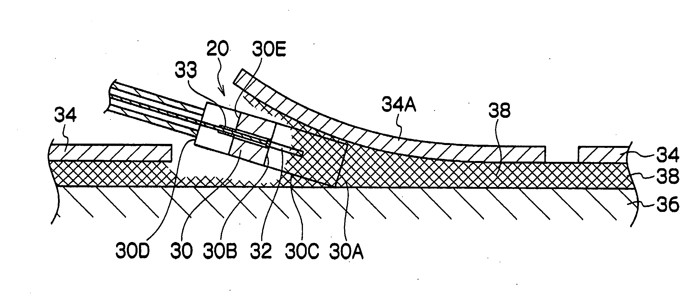
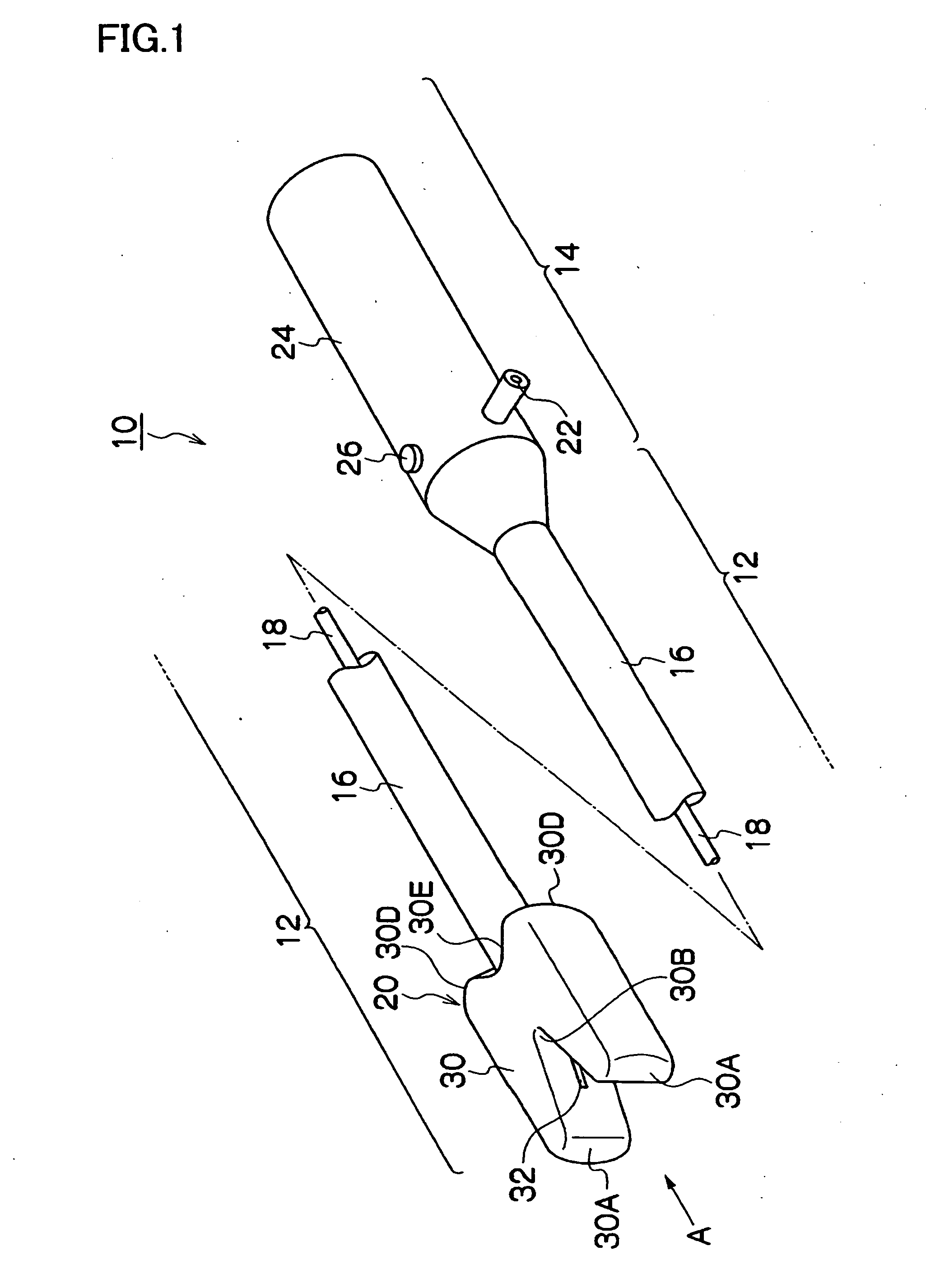
[0061]FIG. 1 is an oblique perspective view illustrating a treatment instrument for an endoscope 10 according to the As shown in the figure, the treatment instrument for an endoscope 10 chiefly comprises an insertion portion 12 that is inserted into a body cavity, and a hand-side operation portion 14 that is provided in a condition in which it is connected with the insertion portion 12. The insertion portion 12 is configured with a non-conductive flexible sheath 16, an electrically conductive wire 18 that is passed through the inside of the flexible sheath 16, and a treatment portion 20 that is attached to the tip of the flexible sheath 16. The tip of the wire 18 is connected to the treatment portion 20, and the proximal end of the wire 18 is connected to a connector 22 of the hand-side operation portion 14. A high-frequency supply apparatus (not shown) that supplies a high frequency current is electrically connected to the connector 22. An operation button 26 is provided on a gras...
second embodiment
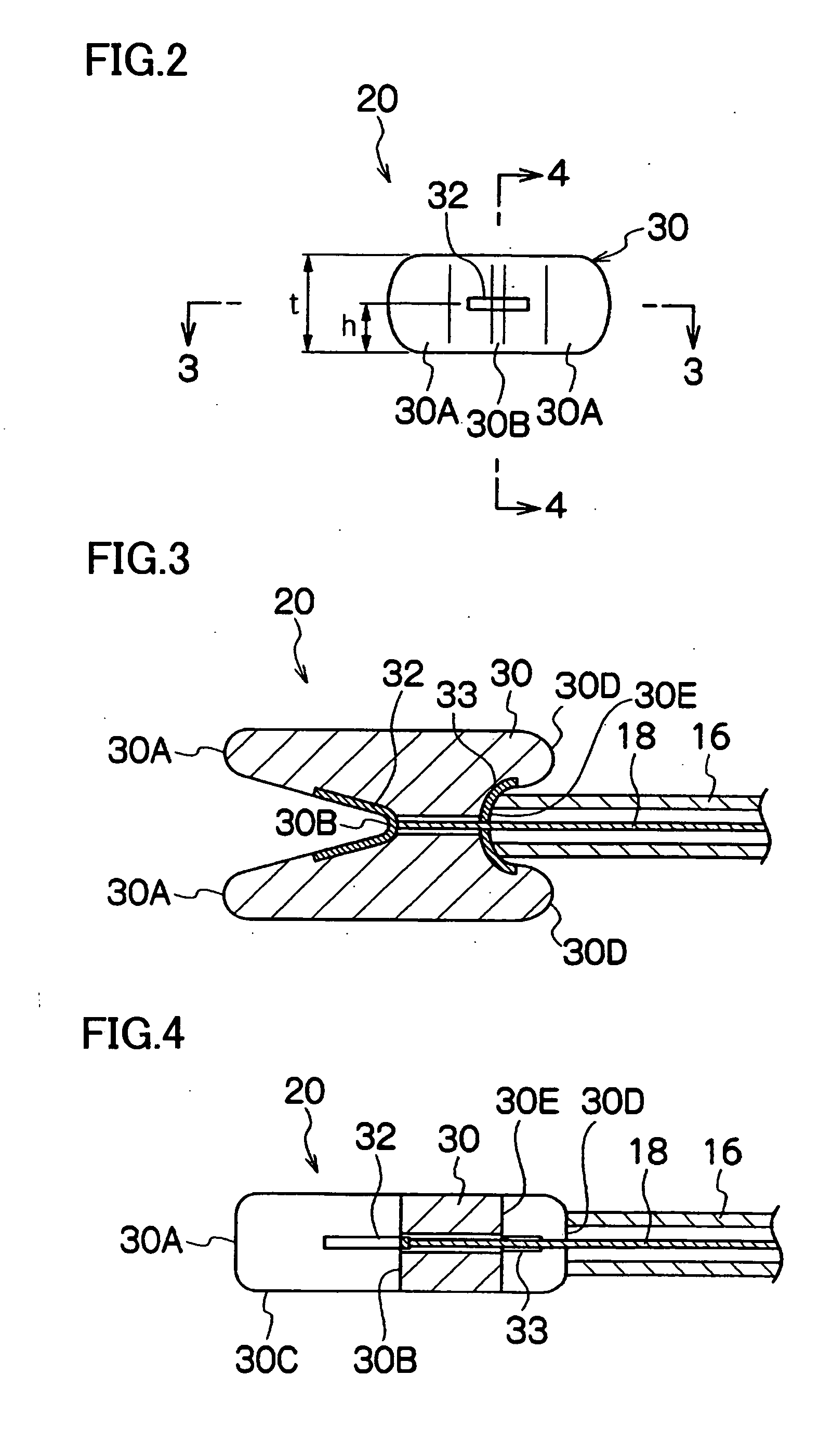
[0080]As shown in these drawings, in a treatment instrument for an endoscope 50 three electrode plates 32, 32, and 32 are provided in a valley portion 30B of a treatment portion 20. The electrode plates 32, 32, and 32 are parallelly disposed at different distances from the underside 30C of the main unit 30. The electrode plates 32, 32, and 32 are respectively connected to different wires 18, 18, and 18, and these three wires 18, 18, and 18 are connected to a changeover switch 52 of a hand-side operation portion 14. The changeover switch 52 is configured to alternatively connect one of the three wires 18, 18, and 18 to the connector 22. Hence, by operating the changeover switch 52, one of the electrode plates 32, 32, and 32 can be selected to feed a high frequency current thereto. The wires 18, 18, and 18 are covered with an outer coat of a non-conductive member or are disposed in a state in which they are separated with a non-conductive partition member so as not to short circuit.
[...
third embodiment
[0084]The treatment instrument for an endoscope is a bipolar type treatment instrument in which a pair of electrodes for feeding a high frequency current is provided in the treatment portion 54. That is, in the treatment portion 54, two electrode plates 32A and 32B are provided in the valley portion 30B of the main unit 30. As shown in FIG. 9, the electrode plates 32A and 32B are disposed at a predetermined distance h from the underside 30C of the main unit 30. Further, the two electrode plates 32A and 32B are, as shown in FIG. 10, opposingly disposed on the sides of the valley portion 30B, and wires 18A and 18B are electrically connected to the electrode plates 32A and 32B. The wires 18A and 18B are connected to the connector 22 of the hand-side operation portion 14 (see FIG. 1). By connecting an unshown high-frequency current supply unit to the connector 22, a high frequency current is passed through the two electrode plates 18A and 18B. The two wires 18A and 18B are covered with...
PUM
 Abstract
Description
Claims
Application Information