
Assessing risk of disease progression in rheumatoid arthritis patients

a rheumatoid arthritis and risk assessment technology, applied in the field of assessing the risk of disease progression in rheumatoid arthritis patients, can solve the problems of loss of function of the involved joints, intense pain and joint destruction, and structural damage progressing

Active Publication Date: 2009-10-29
ROCHE DIAGNOSTICS OPERATIONS INC
View PDF6 Cites 15 Cited by
  • Summary
  • Abstract
  • Description
  • Claims
  • Application Information

AI Technical Summary

Benefits of technology

[0035]Also disclosed is a protein array device comprising at least the appropriate specific binding partners for measurement of CRP and interleukin-6 and optionally appropriate specific binding partners for one or more other marker useful in assessing the risk of disease progression for a patient having rheumatoid arthritis.

Problems solved by technology

As the disease progresses, any type of motion becomes very painful and difficult leading eventually to a loss of function of the involved joints.
The more severe cases of rheumatoid arthritis can lead to intense pain and joint destruction.
Although synovitis tends to follow a fluctuating pattern, structural damage progresses as a linear function of the amount of prior synovitis.
All efforts to associate an infectious agent with RA by isolation, electron microscopy, or molecular biology have failed.
The use of steroids and immunosuppressants, however, has significant risks and side effects both in terms of toxicity and vulnerability to potentially lethal conditions.
Though very promising, biologicals are still in limited use due to high costs.
In all other cases correct diagnosis can be very difficult, especially when the etiology of the disease is not fully understood as is the case for RA.
As discussed above, establishing a diagnosis of RA and deciding for the optimal treatment option is not an easy task.
No unique and generally accepted set of indicators for poor outcome in RA exists to date.
To make things even more complicated, assessing a prognosis in RA also suffers from a lack of a clear and generally accepted definition of disease progression.
However, most of these scores are used in clinical trials settings only, but rarely or not at all in rheumatology practices.
However, the above mentioned tools are far from optimal.
They are time-consuming and influenced by subjective assessments, e.g. in case of the HAQ or tender / swollen joint counts.
However, correlations were not very strong and only existing for the first 6 months of follow-up.
They demonstrated that the determination of anti-CCP at baseline is not a sufficient predictor of disease progression.
Whereas both RF and anti-CCP are important tools in establishing the diagnosis of RA, they appear to be not a strong aid in predicting the future course of disease.
Many markers or sets of markers have been proposed, however the odds ratios achieved thus far have not been sufficient or have been based on a too large variety of biochemical and clinical parameters to meet clinical routine requirements.

Method used


Image

  • Assessing risk of disease progression in rheumatoid arthritis patients
  • Assessing risk of disease progression in rheumatoid arthritis patients
  • Assessing risk of disease progression in rheumatoid arthritis patients

Examples


example 1

Study Population

[0114]Samples derived from 237 highly characterized RA patients with maximum disease duration of 15 years were collected in five European centers with a follow-up of one or two years. All individuals were diagnosed as RA-patients according to the 1987 revised criteria for the classification of Rheumatoid Arthritis from the American Rheumatism Association (Arnett, F. C., et al., Arthritis Rheum. 31 (1988) 315-324). All patients were documented with an extensive case report form (=CRF). The CRF included the Health Assessment Questionnaire, the SF36 Questionnaire, swollen and tender joint count, laboratory parameters, clinical history of relevant surgery, medication, co-morbidities and medication for co-morbidities. X-rays were taken from hands and feet at baseline, after one and after 2 years following a standardized procedure. Only the baseline samples obtained from the RA-patients were used in the measurement of the different analytes and the corresponding results we...

example 2

Determination of the Sharp-Genant-Scores

[0116]From each patient x-rays were taken from hands and feet at baseline and after one and two years. The conventional film radiographs were sent to Synarc (Synarc GmbH, Hamburg, Germany), where the hard copy films were digitized using a Lumiscan 200 high resolution digitizer. After quality check each image was read by an experienced radiologist and scored according the Genant-modified Sharp scoring.

Morphological Scoring of Radiographs

[0117]Bone erosions and joint space narrowing in the hands and feet were scored according to a Genant-modified Sharp grading scheme as described below. This grading scheme is based on the Genant-modified Sharp scoring technique.

[0118]Erosion Score Fourteen sites in each wrist and hand (four proximal interphalangeal and five metacarpophalangeal joints, the carpometacarpal joint of the thumb, the scaphoid bone, the distal radius and the distal ulna) and six joints in each foot (five metatarsophalangeal joints and ...

example 3

Classification of Patients in RA with Disease Progression and RA with No Disease Progression

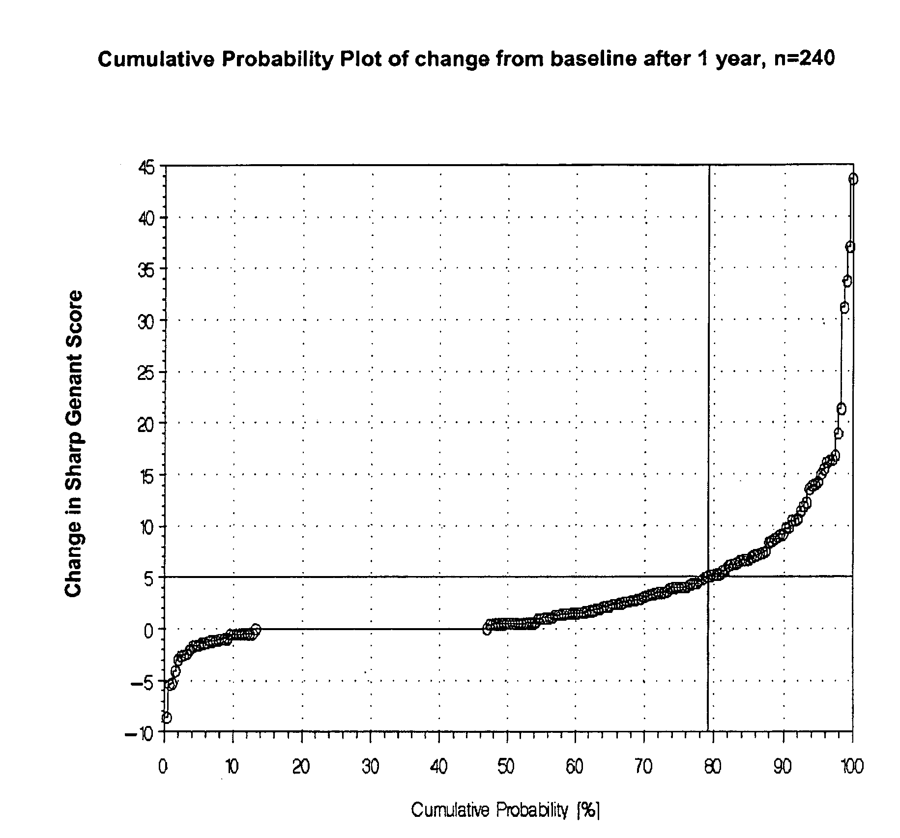
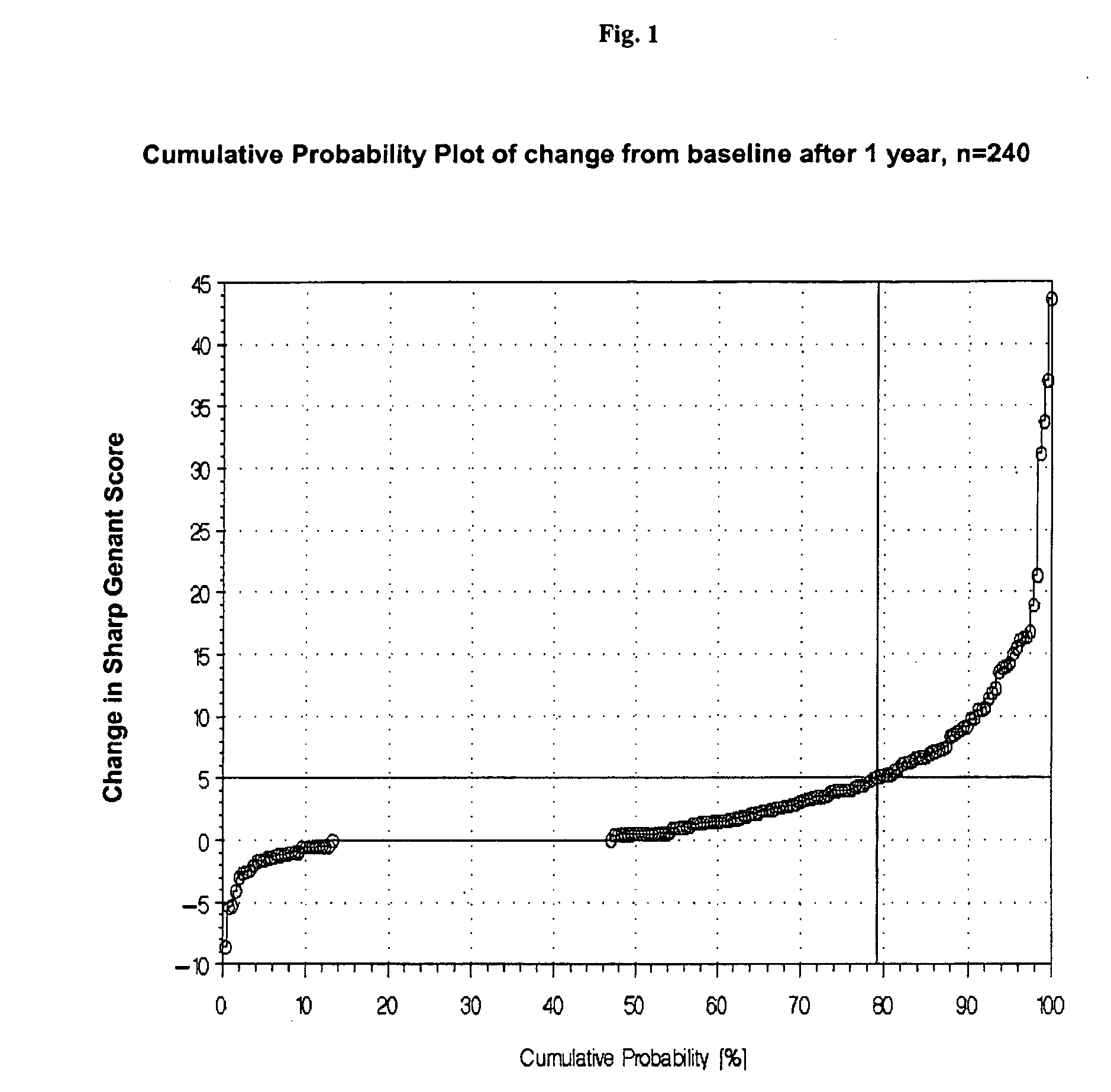
[0125]There are some possibilities discussed in the literature for classification of disease progression. Beside the ACR and EULAR criteria, which are mostly used in pharmaceutical studies for assessing treatment response also the IIAQ score and the radiological scores can be used for classification of disease progression. The most preferred methodology is the use of the change of any radiological score after one year. We decided to use the total Sharp-Genant-Score and to determine the individual change of this score one or two years after the baseline value (=progression rate).

Progression rate (1)=change of Sharp-Genant-Score (SGS) from baseline to year 1.

Progression rate (2)=change of Sharp-Genant-Score (SGS) from baseline to year 2.

[0126]The next important step was to define a cut-off value for the progression rates to be able to classify the patients in RA with progression and RA without ...


PUM

PropertyMeasurementUnit
concentrationaaaaaaaaaa

Abstract

Disclosed is an in vitro method aiding in the further assessment of patients suffering from rheumatoid arthritis. The method especially is used in assessing whether an RA patient is at risk of disease progression. The method is for example practiced by analyzing biochemical markers, comprising measuring in a sample the concentration of at least C-reactive protein (CRP) and interleukin-6 and correlating the concentrations determined to the likelihood of an underlying rapidly progressing form of RA. A patient at high risk of a rapidly progressing disease might be a patient in need for treatment or if already treated in need for a different and more effective treatment. The invention also relates to the use of a marker panel comprising C-reactive protein and interleukin-6 in the assessment of a patient with rheumatoid arthritis and it teaches a protein array device and kit, respectively, for performing the method of the invention.

Description

RELATED APPLICATIONS[0001]This application is a continuation of PCT / EP2007 / 008313 filed Sep. 25, 2007 and claims priority to EP 06020645.5 filed Sep. 29, 2006.FIELD OF THE INVENTION[0002]The present invention relates to an in vitro method aiding in the further assessment of patients suffering from rheumatoid arthritis. The method especially is used in assessing whether an RA patient is at risk of disease progression. The method is for example practiced by analyzing biochemical markers, comprising measuring in a sample the concentration of at least C-reactive protein (CRP) and interleukin-6 and correlating the concentrations determined to the likelihood of an underlying rapidly progressing form of RA. A patient at high risk of a rapidly progressing disease might be a patient in need for treatment or if already treated in need for a different and more effective treatment. The invention also relates to the use of a marker panel comprising C-reactive protein and interleukin-6 in the ass...

Claims


Application Information

Patent Timeline
Patent Type & Authority Applications(United States)
IPC IPC(8): C40B30/04C40B40/10
CPCG01N33/564G01N33/6869G01N2800/102G01N2333/4737G01N2333/5412G01N33/6893
Inventor KARL, JOHANNGRUNERT, VEIT PETERROLLINGER, WOLFGANGWILD, NORBERT
Owner ROCHE DIAGNOSTICS OPERATIONS INC
Learn More