Fluorescent Silica-Based Nanoparticles

a technology of fluorescence silica and nanoparticles, applied in the field of nanoparticles, can solve the problems of substrate-induced changes in either or both the absorption and emission spectrum, difficulty in obtaining highly accurate quantification of cell population's magnetic susceptibility, and general difficulty in accurately determining the chemical and physical structure of cell surface markers and immunological labels attached to the cell surfa

Inactive Publication Date: 2010-02-11
CORNELL RES FOUNDATION INC
View PDF3 Cites 28 Cited by
  • Summary
  • Abstract
  • Description
  • Claims
  • Application Information

AI Technical Summary

Benefits of technology

The patent describes giving patients who need treatment a special type of fluorescent particle that can attach itself to specific cells in the body where the problem is located. This makes it possible for researchers to see those cells glow up when they use certain techniques. The particles may also carry other treatments for the condition.

Problems solved by technology

This patent discusses the uses of fluorescent and magnetic particles in biomedical research and how they can be utilized as markers and indicators of certain events. However, current techniques face limitations in determining the exact structure and composition of these markers due to difficulties in attachment chemistry. Additionally, issues exist with magnetic particles relating to their ability to bind specifically to target cells without causing concomitant effects on neighboring cells. There is also a need for brighter and more easily detected fluorescent nanoparticles for better visualization in biological environments.

Method used


Image

  • Fluorescent Silica-Based Nanoparticles
  • Fluorescent Silica-Based Nanoparticles
  • Fluorescent Silica-Based Nanoparticles

Examples


example 1

[0138]Preparation of Red Core Nanoparticles. 10 mg of tetramethylrhodamine-5-(and-6-)isothiocyanate (TRITC) were dissolved in ethanol. The molar ratio of 3-aminopropyltricthoxysilane (APTS) to TRITC was 50 to 1, with 2 mL of ethanol, per mg of APTS. Following complete dissolution of TRITC in ethanol, APTS was added to the reaction vessel. The reaction was stirred in the dark in the glove box for about 12 hours at room temperature.

example 2

Preparation of Green Core Nanoparticles

[0139]5 mg of Alexa Fluor® 488 C5 Meleimide was dissolved in ethanol. The molar ratio of 3-mercaptopropyltriethoxysilane (MPTS) was 100 to 1, with 2 mL of ethanol per mg of MPTS. Following complete dissolution of TRITC in ethanol, MPTS was added to the reaction vessel and stirred for about 12 hours in darkness at room temperature.

[0140]5 mg of Alexa Fluor® 488 carboxylic acid, succinimidyl ester was dissolved in THF. The molar ratio of 3-Aminopropyltriethoxysilane (APTS) to Alexa Flour® 488 carboxylic acid, succinimidyl ester was 100 to 1, with 2 ml of THF per mg of APTS. Following complete dissolution of Alexa Fluor® 488 carboxylic acid, succinimidyl ester in THF, APTS was added to the reaction vessel and stirred for 12 hours in darkness at room temperature. Due to the minute quantities, the fluorescent dye-core adducts were not isolated from the reaction medium before the silica shell was deposited.

example 3

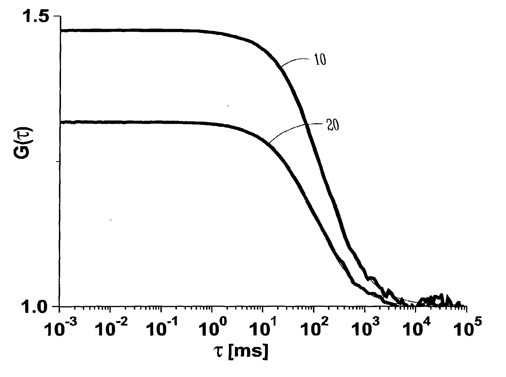
[0141]Preparation of Sized Silica Coated Fluorescent Nanoparticles Preparation of silica coated fluorescent nanoparticles of different sizes was accomplished using different relative molar amounts of reagents as described above. The molar amounts of reagents for the synthesis of 20 nm to 200 nm particles are tabulated in Table I. All silica coating of fluorescent nanoparticle procedures were accomplished at ambient conditions, i.e. at room temperature with ethanol as the solvent. A representative procedure follows.

[0142]To a mixture containing fluorescent core particles there was added continuously 2 mL of TEOS in 100 mL additional solvent, dropwise such as over about 20 minutes. The remaining larger quantity of the TEOS was added at a more rapid rate, such as over about a 45 minute period with simultaneous addition of 400 mL of additional solvent. Optionally, water was added at this stage to grow particles larger than about 100 nm and to maintain the molar ratio of water to TEOS as...


PUM

PropertyMeasurementUnit
Fractionaaaaaaaaaa
Fractionaaaaaaaaaa
Thicknessaaaaaaaaaa

Abstract

The present invention provides nanoparticle compositions comprising, for example, a core comprising a fluorescent silane compound; and a silica shell on the core. Also provided are methods for the preparation of nanoparticle compositions including fluorescent nanoparticles, ligated-fluorescent nanoparticles, ligated-fluorescent nanoparticles having therapeutic agents, and ligated-fluorescent nanoparticles coupled or associated with an analyte. Also provided are methods: for the detection of the ligated-fluorescent nanoparticles; for associating the linked-fluorescent nanoparticles with a cellular component of interest and recording or monitoring the movement of the cellular component; for improving the therapeutic properties of the therapeutic agent by combining the therapeutic agent with linked-fluorescent nanoparticles and contacting or administering the combination to a cell or organism; for making and using the fluorescent nanoparticles in, for example, diagnostic agents for the detection of various analytes, and like applications.

Description


Claims


Application Information

Patent Timeline
Owner CORNELL RES FOUNDATION INC
Why Eureka
Try Eureka