
Medical appliance optical delivery and deployment apparatus and method

a technology for optical delivery and medical appliances, applied in the field of medical devices, can solve the problems of affecting patient ventilation, not giving adequate visual certainty, tissue luminal inflammation and tissue granulation, etc., and achieve the effect of convenient and safe delivery of stents

Active Publication Date: 2010-03-04
MERIT MEDICAL SYST INC
View PDF101 Cites 18 Cited by
  • Summary
  • Abstract
  • Description
  • Claims
  • Application Information

AI Technical Summary

Benefits of technology

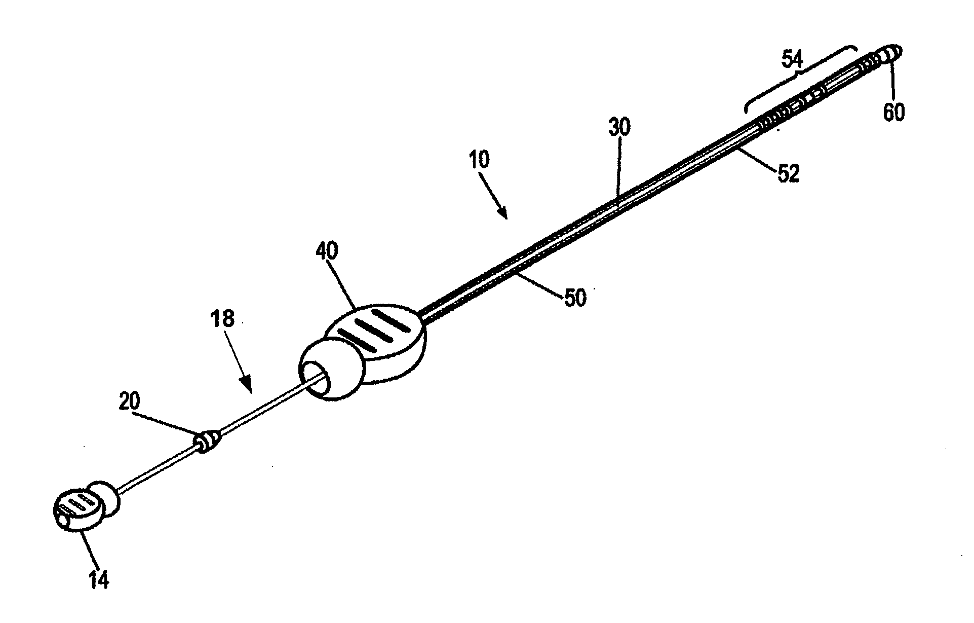
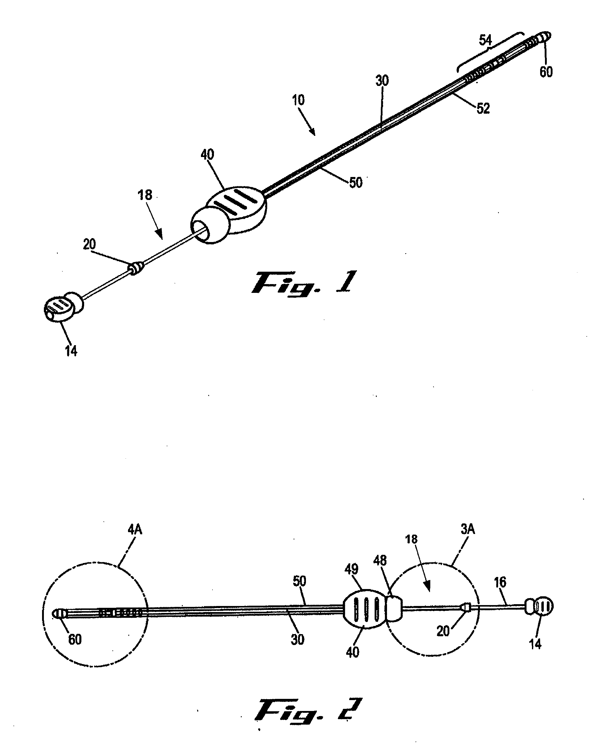
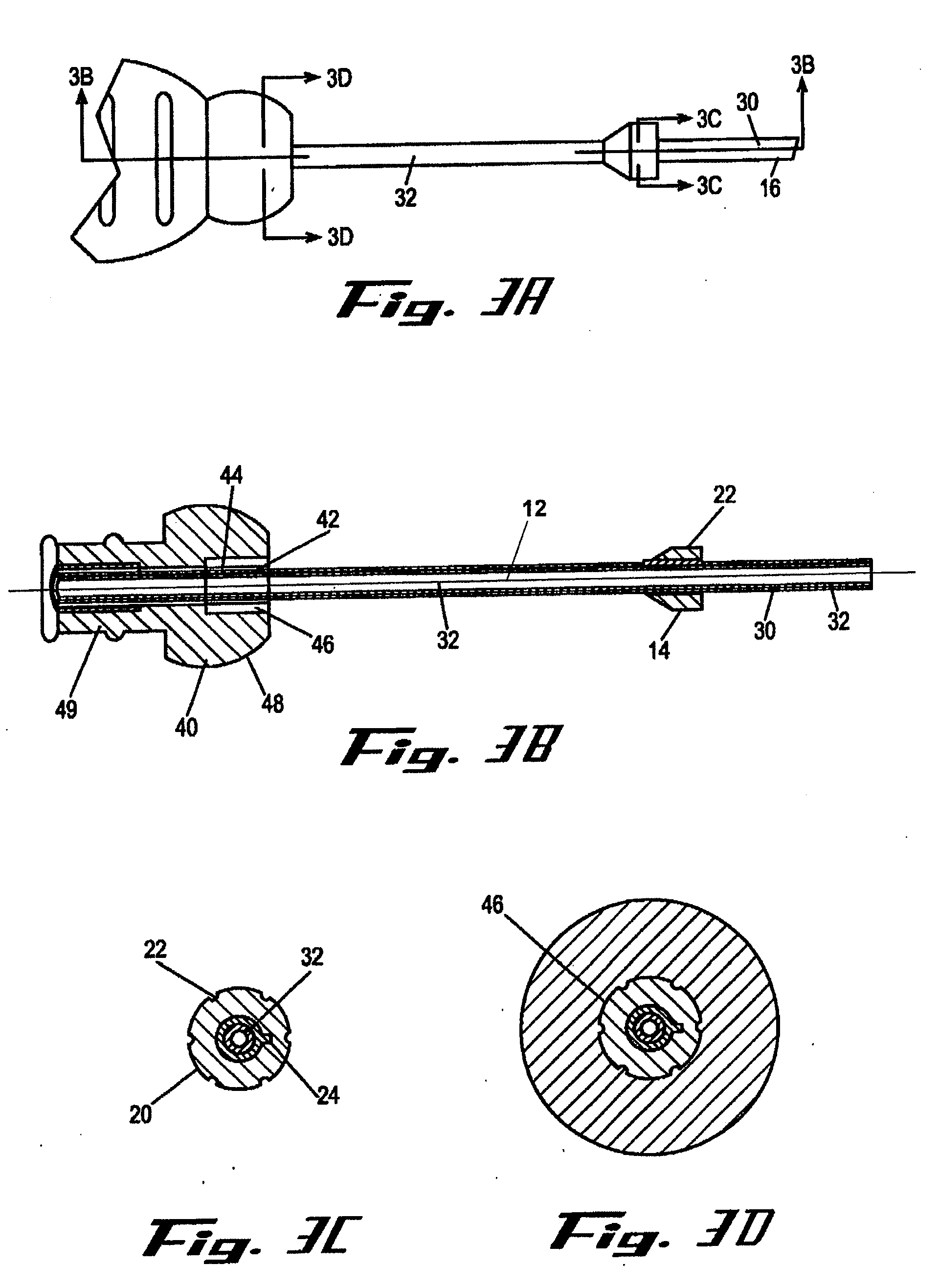
The present invention is a stent deployment apparatus that provides a safer and more precise way to deliver stents in the body. The apparatus allows for easy placement and can be used with various stent designs. It has a smooth and clean outer surface, is flexible and adaptable to different luminai curvatures, and has low friction between the inner and outer tubular members. The apparatus also allows for easy repositioning of the stent and provides direct visualization during the deployment process. It is designed to improve the accuracy and safety of stent deployment and can be used in tracheal respiratory bronchial stenosis.

Problems solved by technology

Though stents are excellent devices when used properly, improper installation can lead to tissue luminal inflammation and tissue granulation.
In particular, many physicians introduce stents with catheters and other delivery devices that do not give them adequate visual certainty that the device has been installed at the desired target site.
Moreover, devices that allow for limited visual feedback have an excessively large diameter, which can hinder patient ventilation.
Additionally, such devices do not have safety features to ensure that the stent is not prematurely and irretrievably deployed.
Unfortunately, guidance of the stent has substantially remained a function of physician skill resulting from substantial practice.
If after full deployment of the stent, the physician discovers the stent has been implanted incorrectly, there is no conventional way of correcting the error short of removing the stent.
In particular, as a rule of thumb, once the exterior catheter, of conventional delivery devices, has been retracted beyond 60%, it generally cannot be realigned with respect to the stent.
Conventional stent delivery devices, however, do not have any safety mechanism to prevent excessive deployment of a misaligned stent.
In fact, conventional delivery devices require the physician to estimate extent of deployment, which results in either overly conservative or excessive deployment—both of which leads to stent misplacement.
As a result visualization features are not directly integrated into the design of these devices and therefore substantially limit their efficacy.
An additional limitation of conventional stent delivery devices is the distal tip of conventional stent delivery devices are not adequately designed to (1) facilitate the clearance of obstructed lumen, or (2) facilitate the removal of the delivery device once the stent is radially expanded.
In particular, most distal tips are not configured to comfortably guide the delivery device through a diseased or occluded lumen so that the stent can be delivered in the most beneficial location.
Moreover, once the stent is radially expanded conventional designs rely exclusively on dimensional mismatching to ensure proper removal of the delivery device.
In the event the stent does not adequately expand to preset dimensions, a conventional delivery device would be stuck in the patient until some invasive procedure is performed to remove it and the defective stent.

Method used


Image

  • Medical appliance optical delivery and deployment apparatus and method
  • Medical appliance optical delivery and deployment apparatus and method
  • Medical appliance optical delivery and deployment apparatus and method

Examples


Embodiment Construction

[0011]It is a principal objective of an exemplary stent deployment apparatus in accordance with the present invention to provide a device that can facilitate the precise delivery of stents in a safe and repeatable fashion. In the furtherance of this and other objectives, a preferred deployment apparatus allows the physician to concentrate on correct placement without having to estimate extent of deployment. In particular, in a preferred embodiment, the present deployment apparatus has a physical safety mechanism that limits deployment to the critical deployment point (i.e., ˜60%). The critical deployment point may range from 5% to 95% but is preferably about 60%. At this point, if the physician is satisfied with placement, she can engage the safety means to what we refer to as the Proceed Orientation (PO) and fully deploy the stent. It is preferred that when the safety mechanism is engaged to the PO, a physical twist and a possible audible indicator sounds to inform the physician th...


PUM


Abstract

Embodiments of the present invention are directed to devices for allowing a user to deploy a stent in an anatomical lumen of a patient. For example, one embodiment is directed to a device including a longitudinally extending inner tubular member and a longitudinally extending outer tubular member that are longitudinally and axially displaceable relative to one another. The outer tubular member includes at least one longitudinally extending channel formed between the exterior and interior diameter of the outer tubular member. In addition, the device includes a handle configured to displace the outer tubular member and inner tubular member relative to each other in response to user intervention and a stop configured to coaxially engage the handle to form a safety mechanism. Displaceability of the outer tubular member and inner tubular member relative to each other is limited by the safety mechanism to a predetermined threshold.

Description

CROSS REFERENCE TO RELATED APPLICATIONS[0001]This patent application is a divisional application of U.S. non-provisional application Ser. No. 10 / 404,197, filed Mar. 31, 2003, which is incorporated in its entirety by this reference.FIELD OF THE INVENTION[0002]The present invention relates generally to medical devices directed to the prevention of nonvascular vessel or passageway occlusion, and more particularly to scent deployment apparatuses and methods for utilizing these devices in the treatment of both benign and malignant conditions.BACKGROUND OF THE INVENTION[0003]Self-expanding stents are valuable prostheses for keeping lumen open and preventing closure due to a stricture, external compression, or internal obstruction. In particular, stents are commonly used to keep blood vessels open in the coronary arteries and they are frequently inserted into the ureters to maintain drainage from the kidneys, the bile duct for pancreatic cancer or cholangiocarcinoma or the esophagus for st...

Claims


Application Information

Patent Timeline
Patent Type & Authority Applications(United States)
IPC IPC(8): A61F2/84A61F2/00A61F2/06
CPCA61F2/95A61F2250/0091A61F2/966
Inventor MANGIARDI, ERIC K.BORG, ULF R.REYNOLDS, JASON M.
Owner MERIT MEDICAL SYST INC
Learn More