Novel fluorescent dyes and compounds, methods and kits useful for identifying specific organelles and regions in cells of interest
a fluorescent dye and compound technology, applied in the field of fluorescent dyes and compounds, can solve the problems of limited scope, fluorescently-labeled antibodies have few practical applications for intracellular imaging in living cells, and the time to market for new drugs is shorter
- Summary
- Abstract
- Description
- Claims
- Application Information
AI Technical Summary
Benefits of technology
Problems solved by technology
Method used
Image

Examples
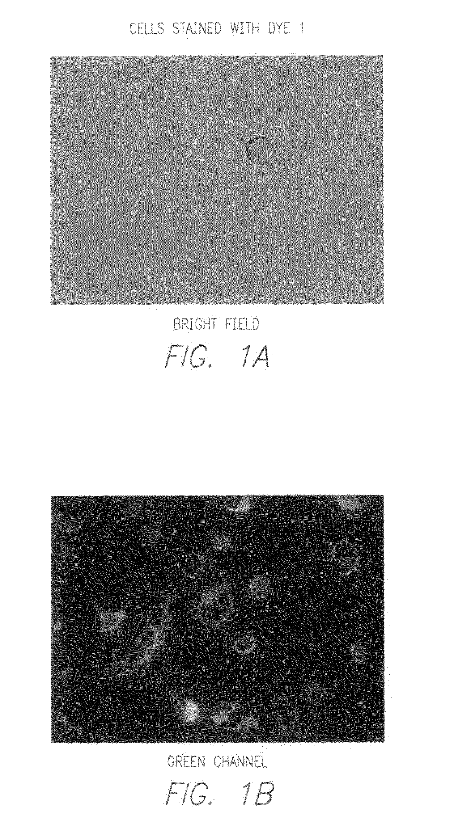
example 1
Synthesis of Dye 1
[0102](a) Preparation of Ethyl 3-(4-methylquinolinium-1-yl) propylphosphonate (Compound 1)
[0103]A mixture of lepidine (1.0 g, 7.0 mmol) and diethyl-(3-bromopropyl)-phosphonate (2.0 g, 7.7 mmol) was heated in a pressure tube at 130° C. for 4 hours. The mixture was allowed to cool to room temperature, and the resulting mass was dissolved in DMF (4 ml). The combined mixture was then added dropwise to ethyl acetate (40 ml). An oily residue was obtained which was washed with ethyl acetate (2×40 ml) and dried under vacuum to yield 1.9 g of Compound 1 which was then used without any further purification. The structure of Compound 1 is given below:
(b) Preparation of Dye 1
[0104]A mixture of Compound 1 (0.274 g, 0.68 mmol), 3,4-bis(benzyloxy)benzaldehyde (0.26 g, 0.82 mmol) and piperidine (33 μL, 0.33 mmol) was refluxed in ethanol (5 ml) using a microwave reactor (CEM, 200 W, open vessel) for 20 mins. The reaction mixture was cooled to room temperature and reaction mixture a...
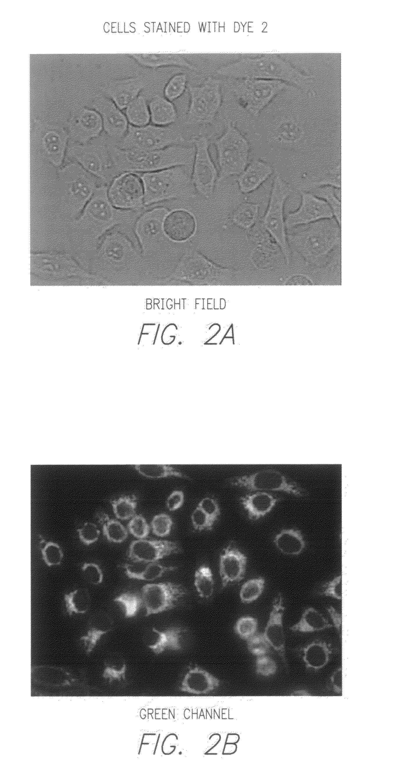
example 2
Synthesis of Dye 2
(a) Preparation of 3,4-dibutoxybenzaldehyde (Compound 2)
[0105]To a solution of 3,4-dihydroxybenzaldehyde (0.5 g, 3.62 mmol) and iodobutane (1.46 g, 7.96 mmol) in ethanol (5 mL), potassium carbonate (1.5 g, 10.9 mmol) was added. Combined mixture was refluxed in the microwave reactor (CEM, 200 W, open vessel) for 1 hour. The reaction mixture was cooled to room temperature, diluted with water (20 mL) and extracted with methylene chloride (2×50 mL). The combined organic layers were washed with water (2×), brine (2×), dried (MgSO4) and evaporated to provide the product as an oily residue (280 mg). This product was used without any further purification. The structure of Compound 2 is given below:
(b) Preparation of Dye 2
[0106]A mixture of Compound 1 (0.2 g, 0.5 mmol), Compound 2 (0.14 g, 0.55 mmol) and piperidine (22 μL, 0.22 mmol) was refluxed in ethanol (4 mL) using a microwave reactor (CEM, 200 W, open vessel) for 1 hour. The reaction mixture was cooled to room tempera...
example 3
Synthesis of Dye 3
(a) Preparation of 1-(3-(diethoxyphosphoryl)propyl)-4-methylpyridinium bromide (Compound 3)
[0107]A mixture of picoline (0.5 g, 5.4 mmol) and diethyl(3-bromopropyl)-phosphonate (1.54 g, 5.9 mmol) was heated in a pressure tube at 130° C. for 4 hours. The mixture was allowed to cool to room temperature, and the resulting mass was dissolved in DMF (4 ml). The combined mixture was then added drop wise to ethyl acetate (40 ml). An oily residue was obtained which was washed with ethyl acetate (2×40 ml) and dried under vacuum to yield 1.7 g of Compound 3 which was then used without any further purification. The structure of Compound 3 is given below:
(b) Preparation of Dye 3
[0108]A mixture of Compound 2 (77 mg, 0.31 mmol), Compound 3 (100 mg, 0.28 mmol) and piperidine (12 μL, 0.13 mmol) was refluxed in ethanol (2 mL) using a microwave reactor (CEM, 200 W, open vessel) for 1 hour. The reaction mixture was cooled to room temperature and solvents were removed in the rotary eva...
PUM
 Abstract
Description
Claims
Application Information
 - R&D Engineer
- R&D Manager
- IP Professional
- Industry Leading Data Capabilities
- Powerful AI technology
- Patent DNA Extraction
Browse by: Latest US Patents, China's latest patents, Technical Efficacy Thesaurus, Application Domain, Technology Topic.
© 2024 PatSnap. All rights reserved.Legal|Privacy policy|Modern Slavery Act Transparency Statement|Sitemap