Device for needle biopsy with integrated needle protection

a technology of endoscope and device, applied in the field of##endoscopic ultrasound procedures, can solve the problems of cumbersome means of attaching a device to an echo-endoscope, inability to examine nearby organs, and inability to perform ovulation diagnostics, etc., and achieve the effect of improving maneuverability and operation of the devi

Active Publication Date: 2010-05-13
TYCO HEALTHCARE GRP LP
View PDF115 Cites 69 Cited by
  • Summary
  • Abstract
  • Description
  • Claims
  • Application Information

AI Technical Summary

Benefits of technology

[0014]According to an aspect of the present disclosure, a device for needle biopsy is presented. The needle biopsy device is comprised of a handle member, a proximal handle member, a distal handle member, a sheath lumen, a needle housing member, a needle, a stylet, and ergonomic design features, including a conical grip, to enhance the maneuverability and operation of the device.
[0017]According to yet another aspect of the present disclosure, a device for needle biopsy is presented. The needle biopsy device is comprised of a handle member, a proximal handle member, a distal handle member, a sheath lumen, a needle housing member, and a needle including a plurality of depressions to enhance echogenicity and ultrasonic visibility.

Problems solved by technology

Although a high frequency ultrasound can produce a higher image quality, high frequency ultrasounds do not penetrate organ walls as well as lower frequency ultrasound.
As a result, the examination of the nearby organs is not possible.
These device, however, suffer from several drawbacks.
For example, the means of attaching a device to an echo-endoscope is cumbersome.
In addition, these devices provide sub-optimal ergonomics of use.
The cumulative actions required by a clinician result in significantly drawn out procedures.
Further, needles commonly kink or deform during removal from a device causing numerous delays and failures.
Moreover, multiple passes per procedure are required, which prolong the procedure and result in a clinician needing to reconfirm the location of a needle relative to a desired aspiration site with each new pass.
Another drawback involving a typical echoendoscope concerns the lack of needle safe preventative design features which protect the clinician from inadvertent needle penetration and the transfer of blood-borne pathogens from a patient to attending medical staff.
As such, the needle sharp of the device is exposed during removal and transfer of the fine needle aspiration device among medical staff in the endoscopic ultrasound and endo-bronchial ultrasound suite with increased risk of “needle sticking” and blood borne pathogen contamination and exposure to same.
Smooth cylindrical surfaces of needles are unfortunately very difficult to image using ultrasonography due to the specular (mirror-like) surface finish of the needle in the untreated state.
Various techniques (sandblasting, surface etching and coating of surfaces) have been used to “roughen” the surface of a needle component with limited success.
This surface roughening results in a scattering of rays from the ultrasound.
However, some of the drawbacks of the aforementioned techniques concern the angle of incidence (sound waves from the ultrasonic transducer) and the angle of reflection (sound waves reflected back to the transducer array).

Method used


Image

  • Device for needle biopsy with integrated needle protection
  • Device for needle biopsy with integrated needle protection
  • Device for needle biopsy with integrated needle protection

Examples


Embodiment Construction

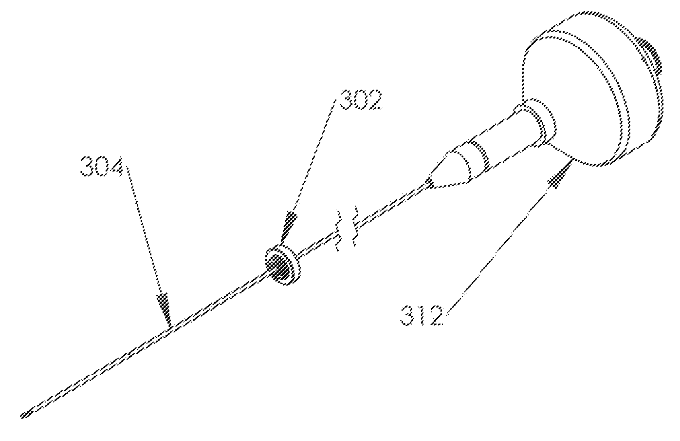
[0084]The exemplary embodiments of the present disclosure are discussed in terms of needle biopsy devices for collecting tissue, fluid, and cell samples from a patient in conjunction with an endoscopic ultrasound or endoscopic bronchial ultrasound. It is contemplated that various embodiments of needle biopsy devices may include a modular design. For example, the needle biopsy device may include a needle housing member that detaches from the proximal handle member of the device for each individual pass or aspirated sample taken by a clinician at the site of lesion or abnormality. In addition, potential design embodiments are disclosed herewith that facilitate needle sharp safety and protection thereof, when combined with devices that incorporate an integrated catheter drive, needle advancement, needle retraction mechanism, and needle in the same device.

[0085]It is envisioned that the present disclosure finds application to a wide variety of biopsy devices for the collection of sample...


PUM


Abstract

A device for needle biopsy is presented. The device includes a handle member having proximal and distal portions. A proximal handle member is disposed to the proximal portion of the handle member and a distal handle member is disposed to the distal portion of the handle member. A sheath lumen is disposed within the handle member and extends from the distal portion of the handle member. A needle housing member is partially disposed to the proximal portion of the handle member and a needle is disposed within the sheath lumen. A plurality of protrusions is disposed upon the needle. A needle protection member is partially disposed to the distal portion of the needle housing member. The needle protection member includes a needle protection hub and a needle protection shaft.

Description

CROSS-REFERENCE TO RELATED APPLICATIONS[0001]This application is a continuation-in-part application of U.S. patent application Ser. No. 12 / 243,367, filed on Oct. 1, 2008, and claims the benefit of U.S. Provisional Application No. 61 / 117,966, filed on Nov. 26, 2008, the entire contents of each are incorporated herein by reference.BACKGROUND[0002]1. Background of Invention[0003]Endoscopic ultrasound procedures have been used for more than twenty five years within the field of medicine. These procedures allow clinicians to scan, locate and identify individual layers of a patient's gastrointestinal tract to determine the location of individual mucosal and sub-mucosal layers. Once identified, appropriate therapeutic modes of treatment for malignancies and various abnormalities may be determined by a clinician.[0004]An endoscopic ultrasound procedure may consist of several steps. For example, a clinician may sedate a patient and insert a probe via esophagogastroduodenoscopy into the patie...

Claims


Application Information

Patent Timeline
Patent Type & Authority Applications(United States)
IPC IPC(8): A61B10/00A61B17/20
CPCA61B10/0283A61B10/04A61B2017/00424A61B2019/5425A61B2017/3413A61B2019/4805A61B2017/00477A61B2090/0801A61B2090/3925A61B10/0266A61B2010/045
Inventor MUGAN, JOHNMURPHY, BRIANMCWEENEY, JOHN
Owner TYCO HEALTHCARE GRP LP
Why Eureka
Try Eureka