Apparatus and method of optical imaging for medical diagnosis

a technology of optical imaging and medical diagnosis, applied in the field of three-dimensional imaging, can solve the problem of few feature points, and achieve the effect of reducing the complexity of the 3-d surface reconstruction problem and improving the shape reconstruction

Active Publication Date: 2010-06-17
THE HONG KONG UNIV OF SCI & TECH
View PDF6 Cites 104 Cited by
  • Summary
  • Abstract
  • Description
  • Claims
  • Application Information

AI Technical Summary

Benefits of technology

[0015]This system significantly reduces the complexity of the 3-D surface reconstruction problem and particularly improves the shape reconstruction of objects having plain spatial characteristics such as human tissue. The imaging method and system can be used for reconstructing the surface of objects such as the cervix with relatively plain spatial characteristics. However, one skilled in the art will understand, after reading the description, that the system and method described herein can readily be used for a wide array of applications in addition to colposcopy, including lesion examinations of skin, oral cavities, and internal organs such as the esophagus.

Problems solved by technology

The difficulty in passive stereo vision systems for 3-D sensing includes the problem of occlusion and that very few feature points are available when imaging an object having a smooth surface, such as the human cervix.

Method used


Image

  • Apparatus and method of optical imaging for medical diagnosis
  • Apparatus and method of optical imaging for medical diagnosis
  • Apparatus and method of optical imaging for medical diagnosis

Examples


Embodiment Construction

System Overview

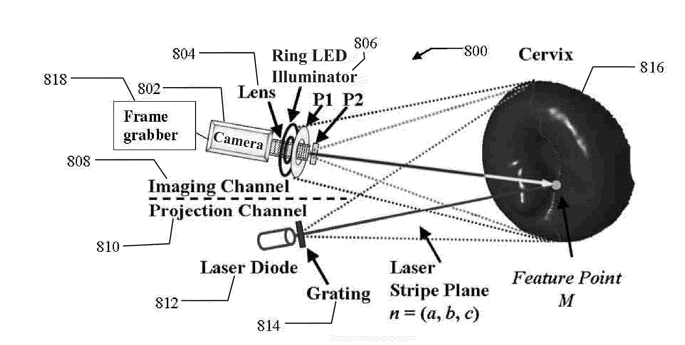
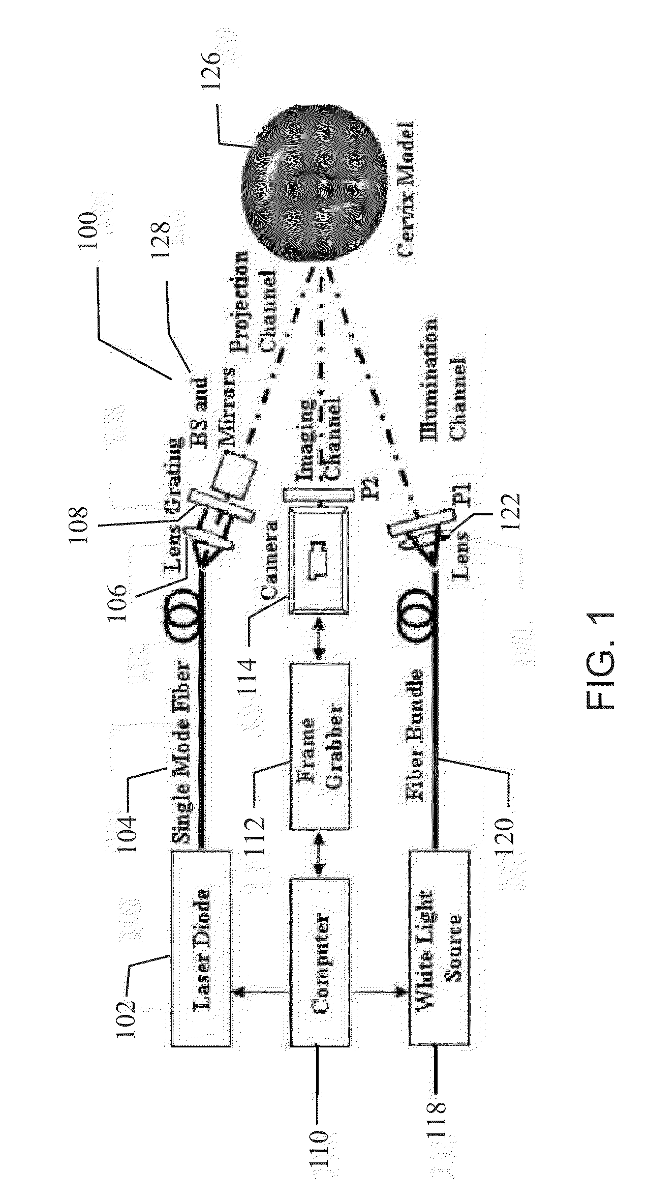
[0059]According to one embodiment illustrated in FIG. 1, a system 100 for providing optical imaging for medical diagnosis includes an imaging channel, an illumination channel, and a projection channel. The imaging channel has the similar configuration as a standard colposcope or a low power microscope. A three-CCD color camera 114 and a fast frame grabber 112 are used to capture images with the resolution of 768×576 pixels in the imaging channel. Two polarizers, P1 and P2, with cross-polarization configuration are placed in front of the camera 114 and the white-light illumination source 118, respectively. This arrangement effectively eliminates the specular reflection from the surface of the examined object 126, which is a human cervix model in this exemplary embodiment. The size and shape of the model are similar to a real human cervix. Landmarks indicated by dots on the model surface are used to evaluate the performance of the imaging system in three-dimensional rec...


PUM


Abstract

Described herein is a novel 3-D optical imaging system based on active stereo vision and motion tracking for to tracking the motion of patient and for registering the time-sequenced images of suspicious lesions recorded during endoscopic or colposcopic examinations. The system quantifies the acetic acid induced optical signals associated with early cancer development. The system includes at least one illuminating light source for generating light illuminating a portion of an object, at least one structured light source for projecting a structured light pattern on the portion of the object, at least one camera for imaging the portion of the object and the structured light pattern, and means for generating a quantitative measurement of an acetic acid-induced change of the portion of the object.

Description

TECHNICAL FIELD[0001]The invention relates to three-dimensional imaging and, in particular, to three-dimensional colposcopy imaging systems.BACKGROUND OF THE INVENTION[0002]Colposcopy is a standard technique to detect early neoplastic growth in cervical tissue. It is a medical diagnostic procedure to examine an illuminated, magnified view of the cervix and the tissues of the vagina and vulva. Many premalignant lesions and malignant lesions in these areas have discernible characteristics which can be detected through this examination technique. It is done using a colposcope, which provides an enlarged view of the areas under examination, allowing the colposcopist to visually distinguish normal from abnormal appearing tissue and take directed biopsies for further pathological examination. The main goal of colposcopy is to prevent cervical cancer by detecting precancerous lesions early and treating them.[0003]Cervical intraepithelial neoplasia (CIN), also known as cervical dysplasia, i...

Claims


Application Information

Patent Timeline
Patent Type & Authority Applications(United States)
IPC IPC(8): H04N13/02H04N7/18H04N5/89G06K9/00G06V10/145
CPCA61B1/00193A61B1/042G06K9/6247A61B5/1076G06K9/2036A61B1/303G06V10/145A61B1/00194A61B1/0605G06V10/7715G06F18/2135
Inventor QU, JIANANWU, TAO
Owner THE HONG KONG UNIV OF SCI & TECH
Why Eureka
Try Eureka