Use of vascular endothelial growth factor receptor 1+ cells in treating and monitoring cancer and in screening for chemotherapeutics

a technology of vascular endothelial growth factor and vascular endothelial cells, which is applied in the direction of antibody medical ingredients, drug compositions, instruments, etc., can solve the problem that none of these studies have shown whether the expression of vegf is associated with parallel surface vegfr2/, and achieve the effect of preventing tumor spread and enhancing tumor cell migration

Active Publication Date: 2010-06-17
CORNELL RES FOUNDATION INC
View PDF5 Cites 4 Cited by
  • Summary
  • Abstract
  • Description
  • Claims
  • Application Information

AI Technical Summary

Benefits of technology

[0022]At present, it is very difficult to determine those patients with solid tumors after complete resection or partial resection and adjuvant chemotherapy that will then develop metastasis. The system of measuring VEGFR1+ bone marrow derived cells in frequent sites of metastasis for any given tumor (for example liver in colon cancer patients, lung in osteosarcoma patients, lymph node in breast cancer patients) can serve to help predict which patients will require additional adjuvant treatment targeted against these VEGFR1+ bone marrow derived clusters as well as more conventional therapy for example upfront treatment after resection with chemotherapy rather than waiting for the presentation of metastatic or recurrent disease. These cellular clusters which may also be tracked in the bloodstream as circulating or mobilized cells expressing VEGFR1 or VLA. In addition, it is possible in the future that these VEGFR1+ hematopoietic progenitor cells may be tagged so as to be followed by imaging studies to follow response to therapy or to assess future metastatic risk. It can serve to change the approach to staging and monitoring for minimal residual disease. Such a marker and potential treatment strategy should prove to be very useful for the care of patients with a primary tumor as well as those patients. It is foreseeable that this strategy may also work for those patients with ischemic disease and inflammatory illnesses such as rheumatoid arthritis and inflammatory bowel disease.
[0023]In the presence of these VEGFR1+ bone marrow-derived cells in sites of metastasis prior to tumor spread leading to the formation of a conducive microenvironment for incoming VEGFR2+ endothelial cells and tumor cells provides evidence that these cells produce a novel chemokine or growth factor which can explain these characteristics. The isolation / identification of novel proteins / genes involved in VEGFR1+ cells and the pre-metastatic niche causing enhanced tumor cell migration and adherence will provide an important target for the prevention of tumor spread.

Problems solved by technology

However, none of these studies have shown whether expression of VEGF is associated with any parallel surface VEGFR2 / VEGFR1 expression or functional response.

Method used


Image

  • Use of vascular endothelial growth factor receptor 1+ cells in treating and monitoring cancer and in screening for chemotherapeutics
  • Use of vascular endothelial growth factor receptor 1+ cells in treating and monitoring cancer and in screening for chemotherapeutics
  • Use of vascular endothelial growth factor receptor 1+ cells in treating and monitoring cancer and in screening for chemotherapeutics

Examples


example 1

Bone Marrow BM Transplantation

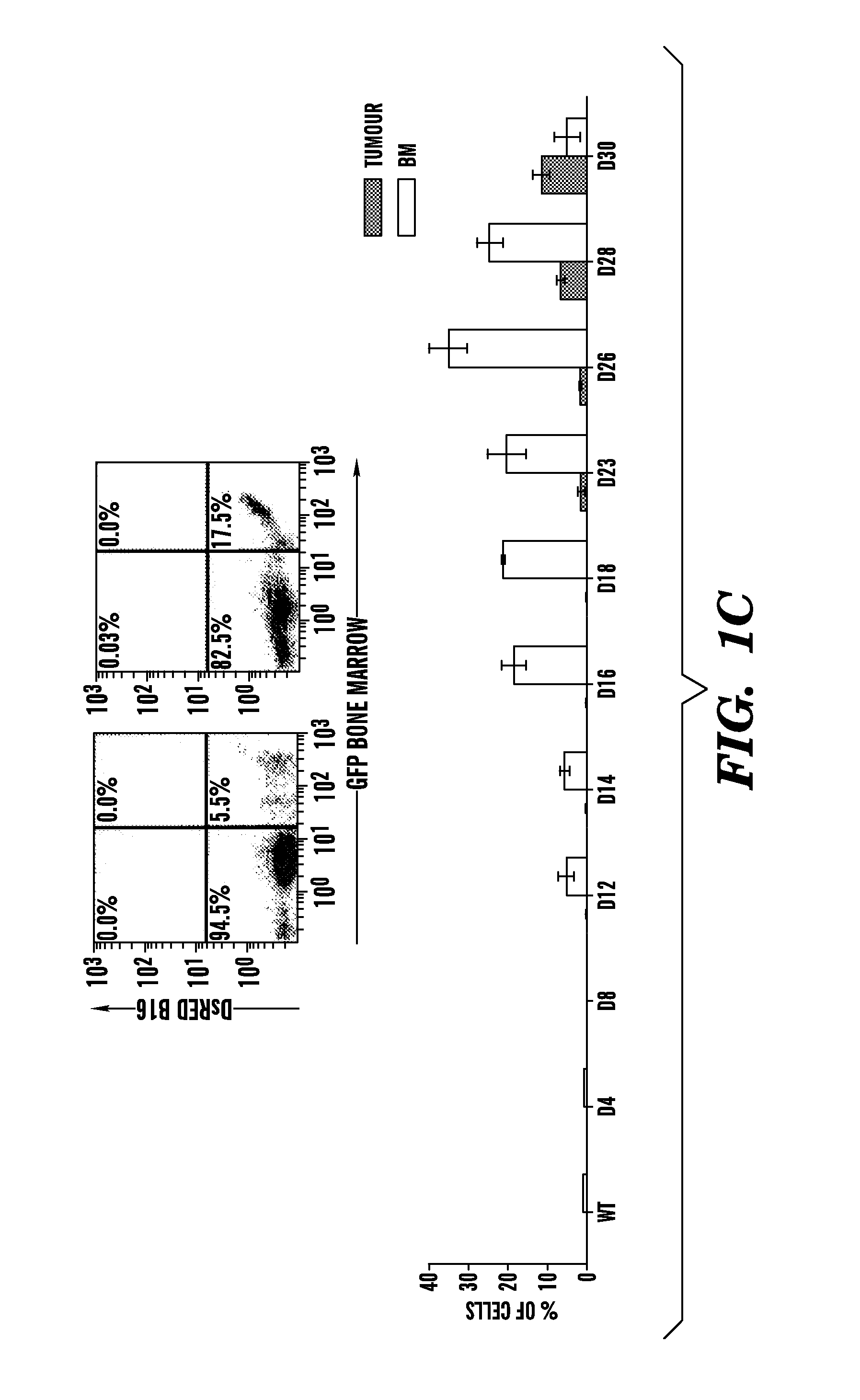
[0087]Wild-type C57B1 / 6 mice were lethally irradiated (950 rads) and transplanted with 1×10⁶ β-galactosidase+ or GFP+ BM cells (Rosa-26 mice) (Lyden et al., “Impaired Recruitment of Bone-marrow-derived Endothelial and Hematopoietic Precursor Cells Blocks Tumor Angiogenesis and Growth,”Nat. Med. 7:1194-1201 (2001), which is hereby incorporated by reference in its entirety). After 4 weeks, mice were injected intradermally with either 2×10⁶ LLC (ATCC) or B-16 cells (ATCC).

example 2

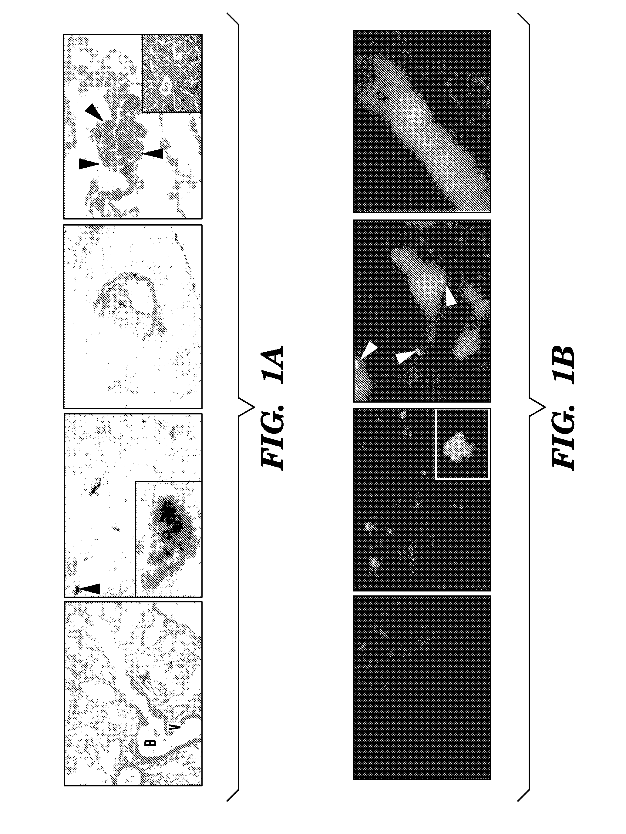
β-Galactosidase Staining

[0088]Tissues and femoral bones were fixed in 4% paraformaldehyde (PFA) for 4 h. The samples were stained in X-gal solution at 37° C. for 36 hours, as described in Tam et al., “The Allocation of Epiblast Cells to the Embryonic Heart and other Mesodermal Lineages The Role of Ingression and Tissue Movement during Gastrulation,”Development. 124:1631-1642 (1999), which is hereby incorporated by reference in its entirety. The X-gal stained tumors and BM were embedded as described in Lyden et al., “Impaired Recruitment of Bone-marrow-derived Endothelial and Hematopoietic Precursor Cells Blocks Tumor Angiogenesis and Growth,”Nat. Med. 7:1194-1201 (2001), which is hereby incorporated by reference in its entirety.

example 3

Fluorescent Tumor Transfection

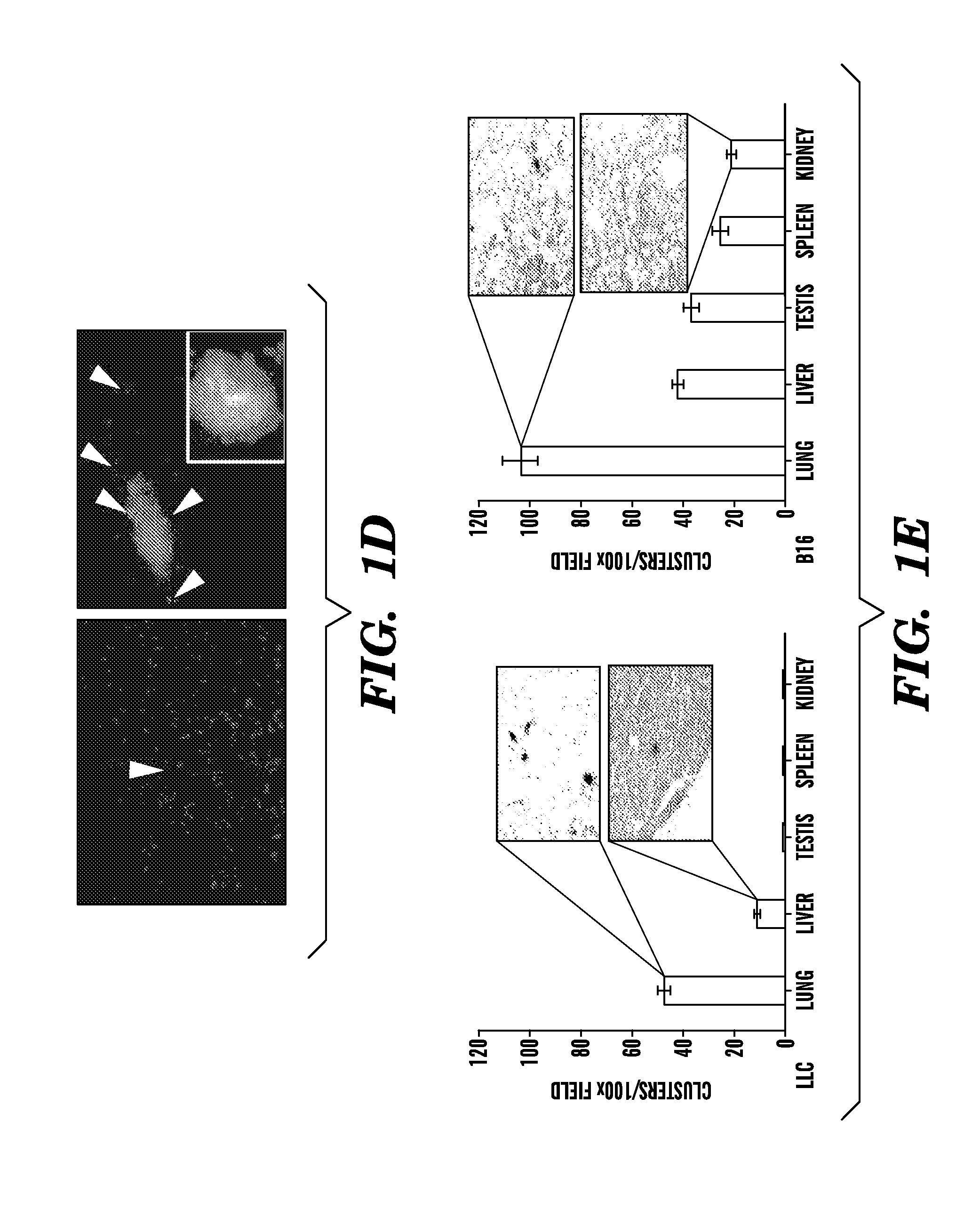
[0089]B16 and LLC cells (2×10⁵) were plated 24 hours prior to transfection. Cells were then pelleted and resuspended into serum free media with lentiviral vector supernatant containing the DsRed reporter gene or GFP gene. Concentrated viral constructs with titers of 1-2×10⁸ infectious particles per ml were used to infect 2×10⁶ cells in a 1 ml (multiplicity of infection is 50) as previously described. Flow cytometric analysis was performed to confirm degree of fluorescence. C57B1 / 6 mice were inoculated with 2×10⁶ LLC / GFP+ or B16 / DsRed+ cells.


PUM


Abstract

The present invention is directed to a method of inhibiting tumor formation in a cancer patient at a site remote from sites of prior tumor formation and to a method of preventing metastases. These methods involve administering to the cancer patient an inhibitor of vascular endothelial growth factor receptor 1+ bone marrow-derived cells under conditions effective either to inhibit tumor formation in the cancer patient at a site remote from sites of prior tumor formation or to prevent metastases. Candidate compounds useful for such purposes can be screened depending on whether they bind to vascular endothelial growth factor receptor 1+ bone marrow-derived cells. Metastases in a cancer patient can be monitored by evaluating a patient sample for detection and quantification of vascular endothelial growth factor receptor 1+ bone marrow-derived cells and comparing the level of vascular endothelial growth factor receptor 1+ bone marrow-derived cells to prior levels.

Description

[0001]This application is a division of U.S. patent application Ser. No. 11 / 284,265, filed Nov. 21, 2005, which claims the benefit of U.S. Provisional Patent Application Ser. Nos. 60 / 629,662, filed Nov. 19, 2004, and 60 / 723,770, filed Oct. 5, 2005, which are hereby incorporated in their entirety.[0002]This invention was made with government support under grant number 1 R01 CA098234-01 awarded by National Institutes of Health. The government has certain rights in this invention.FIELD OF THE INVENTION[0003]The present invention is directed to the use of vascular endothelial growth factor receptor 1+ cells in treating and monitoring cancer and in screening for chemotherapeutics.BACKGROUND OF THE INVENTION[0004]Cancer is the second leading cause of death next to heart attacks in the United States. There has been important progress in the development of new therapies in the treatment of this devastating disease. Much of the progress is due to better understanding of cell proliferation in...

Claims


Application Information

Patent Timeline
Patent Type & Authority Applications(United States)
IPC IPC(8): A61K39/395A61P35/00A61K39/00
CPCA61K2039/505C07K16/2842C07K16/2863G01N2333/71G01N33/5011G01N33/5044C07K2316/96C07K2317/76A61P35/00
Inventor LYDEN, DAVIDKAPLAN, ROSANDRA N.RIBA, REBECCA D.RAFII, SHAHIN
Owner CORNELL RES FOUNDATION INC
Why Eureka
Try Eureka