Enhancement of angiogenesis to grafts using cells engineered to produce growth factors

Active Publication Date: 2005-01-06
WAKE FOREST UNIVERSITY
View PDF22 Cites 108 Cited by
  • Summary
  • Abstract
  • Description
  • Claims
  • Application Information

AI Technical Summary

Benefits of technology

In one preferred embodiment, the matrices are miniaturized for greater ease of implantation. In many applications mini-matrices are desirable having sizes in which the greatest dimension is on the order of 50 millimeters or less, preferably 25 millimeters or less, and most preferably 10 millimeters or less. For example, polymeric wafers can have dimensions of about 2-5 mm, preferably about 2-3 mm. The wafers preferably are thin enough to allow vascularity to occur between the cells on the wafers and those of the surroundings. In one embodiment, the matrix can be seeded with genetically engineered cells capable of stably or transiently producing growth factors or angiogenesis modulating agents.
During in vitro growth, the cells develop and produce a tissue layer which envelopes the matrix material. The tissue layer is capable

Problems solved by technology

Organ transplantation yields a high risk of rejection, even with a good histocompatibility match.
However, these immunosuppressive drugs have a narrow therapeutic window between adequate immunosuppression and toxicity.
Prolonged immunosuppression can weaken immune systems, which can lead to a threat of infections developing

Method used


Image

  • Enhancement of angiogenesis to grafts using cells engineered to produce growth factors
  • Enhancement of angiogenesis to grafts using cells engineered to produce growth factors
  • Enhancement of angiogenesis to grafts using cells engineered to produce growth factors

Examples


example 1

VEGF Secretion from Encapsulated CHO VEGF Cells

I. Material and Methods

Matrix:

A biological acellular collagen matrix was isolated from Swine kidneys. Swine kidneys were harvested, agitated in triton 100X (Sigma St. Louis, Mo.) and ammonium hydroxide (Sigma). The matrices were washed extensively with saline, and allowed to dry. The matrices were lyophilized and cut into 3×3 mm sections. Scanning electron microscopy confirmed the acellular nature of the collagen matrices.

Renal Cells:

Kidneys from 5 day old C57 black mice (Jackson labs; Bar Harbore, Me.) were harvested, minced, suspended in Keratinocyte SFM medium (Gibco BRL; New York, N.Y.) containing Collagenase / Dispase 1 mg / ml (Boehringer Mannheim; Mannheim, Germany) and agitated for one hour at 37° C. The mixed culture of renal cells was filtered serially through 104 μm sieves until all undigested tissue was removed. The filtrate was washed twice with medium and seeded on the collagen matrices at a density of 30×106 cells / m...

example 2

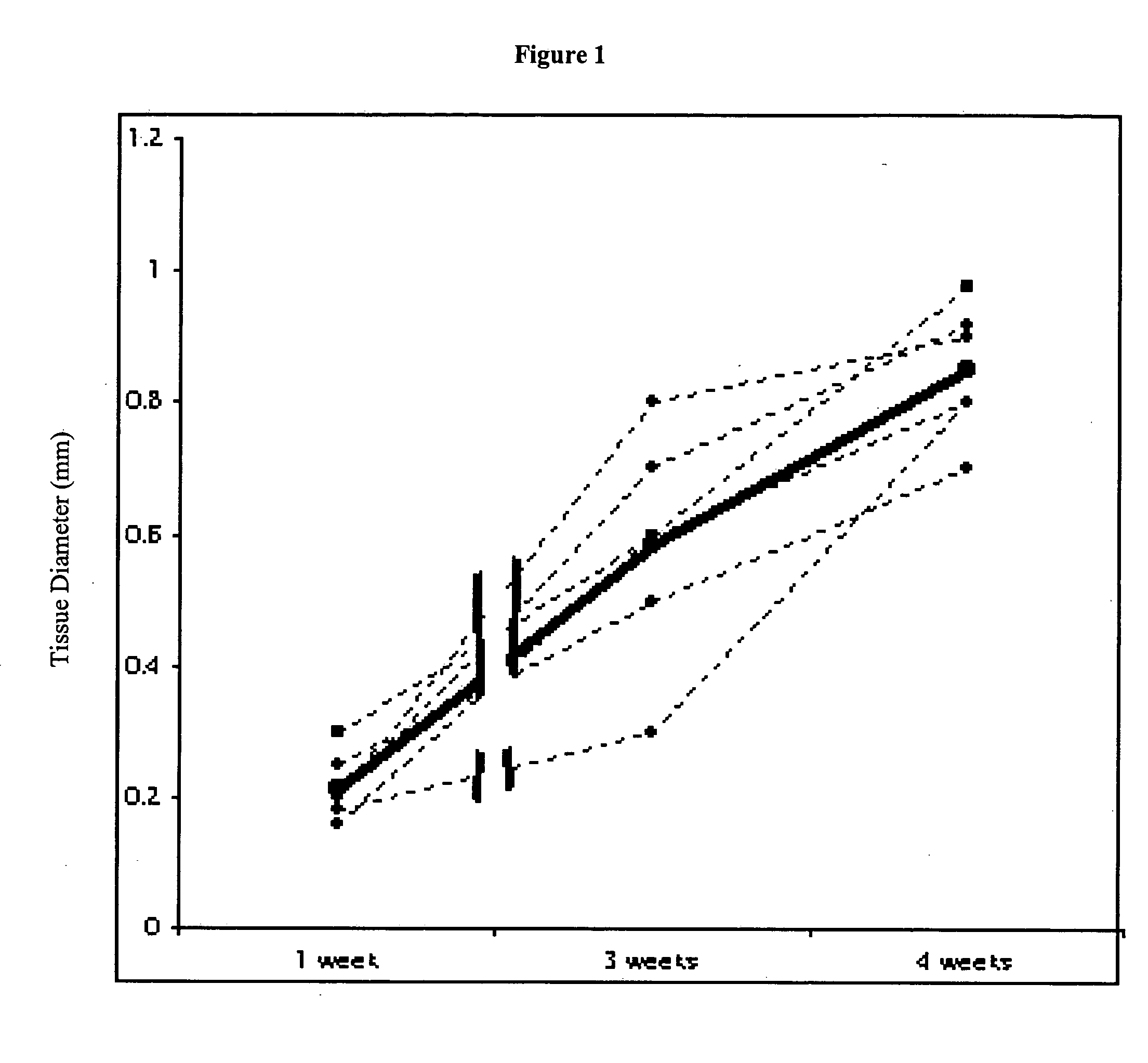
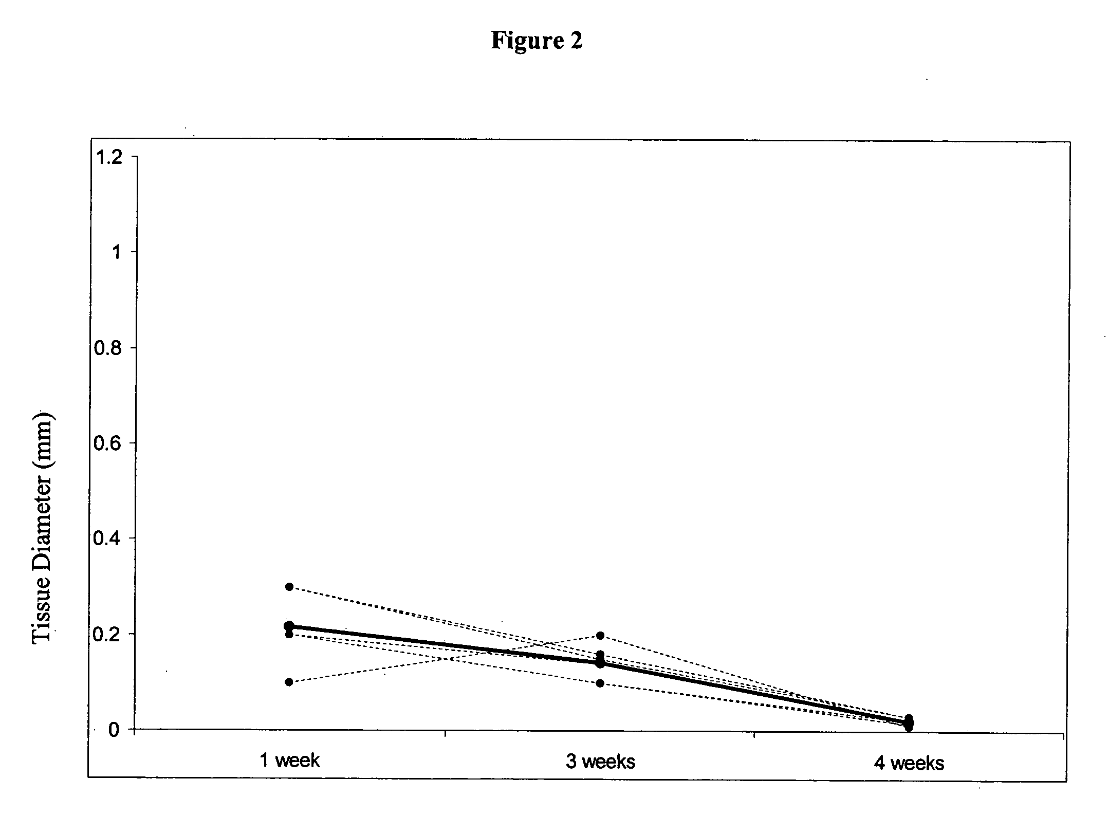
Genetically Engineering Muscle Cells to Transiently Produce VEGF

This study demonstrates that in vivo engineered muscle tissues improve their vascularity when VEGF gene modified muscle cells are used. Desmin positive myoblasts were obtained that formed spontaneously contractile myofibers by 2 weeks in vitro. VEGF and GFP expression in vitro was respectively assessed by Western blot analysis and fluorescence, showing that the myoblasts were successfully transfected. Analysis of the in vivo recovered engineered tissues with RT-PCR and fluorescence showed expression of VEGF and GFP proteins. Immunohistochemical analysis of the recovered tissues confirmed the muscle phenotype. Neovascularization and muscle tissue mass significantly increased with the functional VEGF-transfected cells compared with the non-functional VEGF-transfected cells.

Myoblasts Isolation:

All procedures were done in according with Children's Hospital Animal Care Committee protocols. Myoblasts were derived from s...

example 3

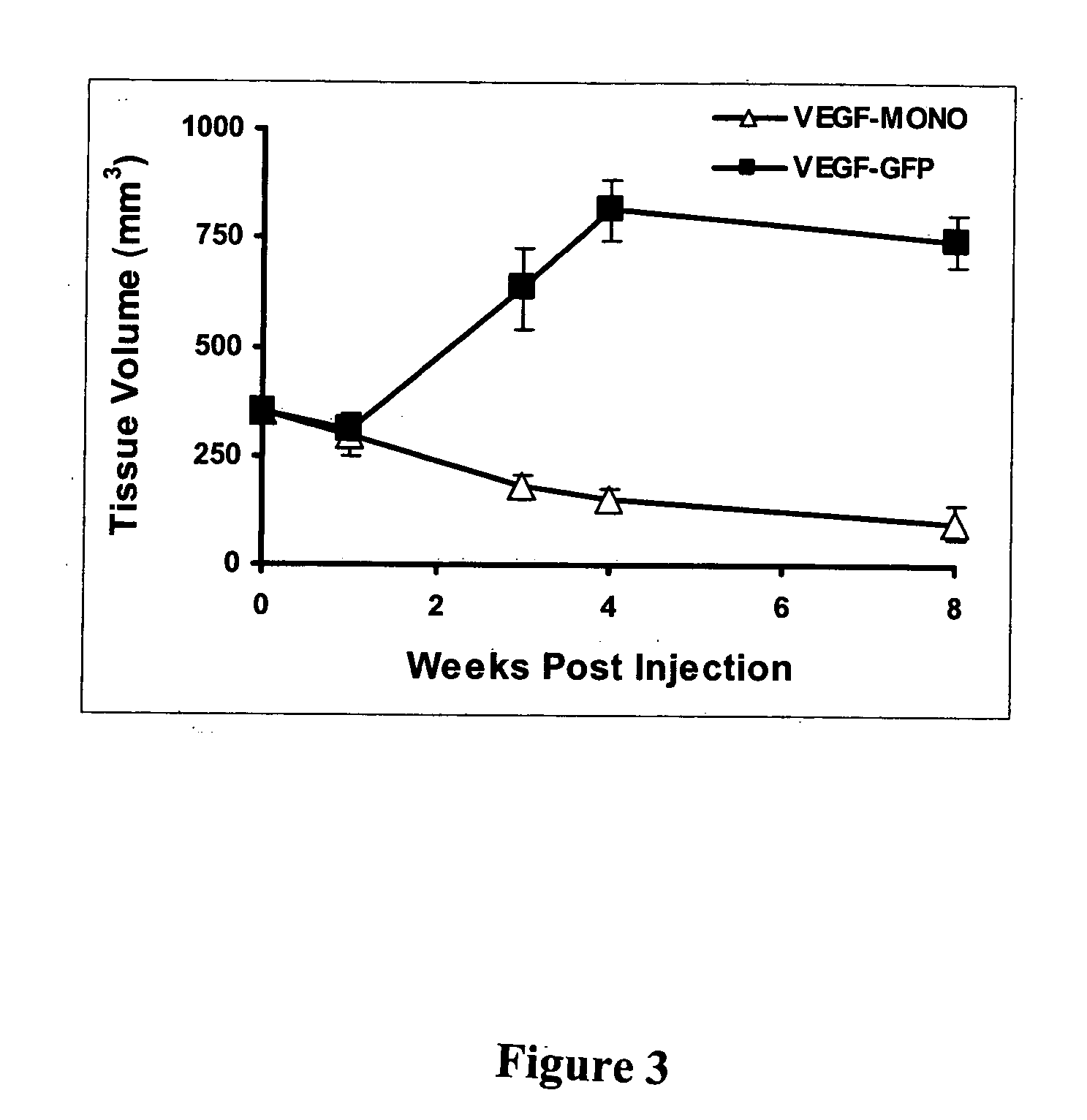
Muscle Formation, Neovascularization, and Volume Preservation in Engineered Tissues Using a Combination of VEGF and Endothelial Cells

This Example illustrates the methods of this invention by demonstrating tissue neovascularization following the implantation of myoblasts and vascular EC in bioengineered tissues. Dermal capillary-derived EC were used in this Example because they represent the same type of the EC in the mouse skin blood vessels that serve the source of EC for subcutaneous neovascularization. Incorporation of EC into the developing tissue had no major effect on tissue volumes of VEGF expressing and control myoblasts. This finding may be explained by the observation that the transplanted vascular EC were a small fraction of the vascular network of the newly formed tissues and the majority was the host (i.e., mouse) EC. However, incorporation of vascular EC enhanced and prolonged MyoD expression and thus, contributed to muscle differentiation of the engineered tissue. T...


PUM

PropertyMeasurementUnit
Timeaaaaaaaaaa
Vascularizationaaaaaaaaaa

Abstract

The present invention provides methods and compositions of engineered cells for use in the continuous or transient delivery of growth factors and angiogenesis modulating agents, such as vascular endothelial growth factor (VEGF), in conjunction with constructs for replacing or augmenting organ functions. In one aspect of he invention, the genetically engineered cells can be immature cells that are capable of differentiating and assimilating into the target region. The methods of the present invention can be used to enhance vascularization locally at a target site in need of repair, growth, or implantation through the incorporation of autologous cells which have been genetically engineered to secrete a growth factor or angiogenesis modulating agent.

Description

FIELD OF THE INVENTION The present invention concerns genetically engineering cells to secrete growth factors and / or angiogenesis modulating agents and methods of enhancing angiogenesis of implanted organ constructs or implanted cultured cell populations. BACKGROUND OF THE INVENTION Injury, congenital abnormalities, disease and aging result in disorders of organs and tissues. Organ transplantation yields a high risk of rejection, even with a good histocompatibility match. Immunosuppressive drugs such as cyclosporin and FK506 are usually given to the patient to prevent rejection. However, these immunosuppressive drugs have a narrow therapeutic window between adequate immunosuppression and toxicity. Prolonged immunosuppression can weaken immune systems, which can lead to a threat of infections developing. In some instances, even immunosuppression is not enough to prevent organ rejection. Therefore, current reconstruction techniques prefer using native tissues from multiple body site...

Claims


Application Information

Patent Timeline
IPC IPC(8): A61K35/12A61K35/34A61K35/44A61K38/18A61K48/00A61L27/38C12N5/071C12N5/077
CPCC12N2501/165A61K35/44C12N2502/253C12N2502/28C12N2502/99C12N2533/32C12N2533/52C12N2533/54C12N2533/72C12N2533/90A61K35/12A61K48/00A61L27/3808A61L27/3834A61L27/3886C12N5/0658C12N5/0686C12N5/0697A61K38/1866A61K35/34C12N2502/1347
Inventor ATALA, ANTHONYSTOKER, SHAY
Owner WAKE FOREST UNIVERSITY
Why Eureka
Try Eureka