Method for production of dendritic cell
a technology of dendritic cells and dendritic cells, which is applied in the field of dendritic cell production methods, can solve the problems of insufficient therapeutic effect, limited number of dc precursor cells, and limited number of progenitor cells, and achieve the effect of strong ability to induce immunity
- Summary
- Abstract
- Description
- Claims
- Application Information
AI Technical Summary
Benefits of technology
Problems solved by technology
Method used
Image
Examples
example 1
Assessment for Cytokine-Induced Expansion and Differentiation of Dendritic Cell (DC) Precursor Cells
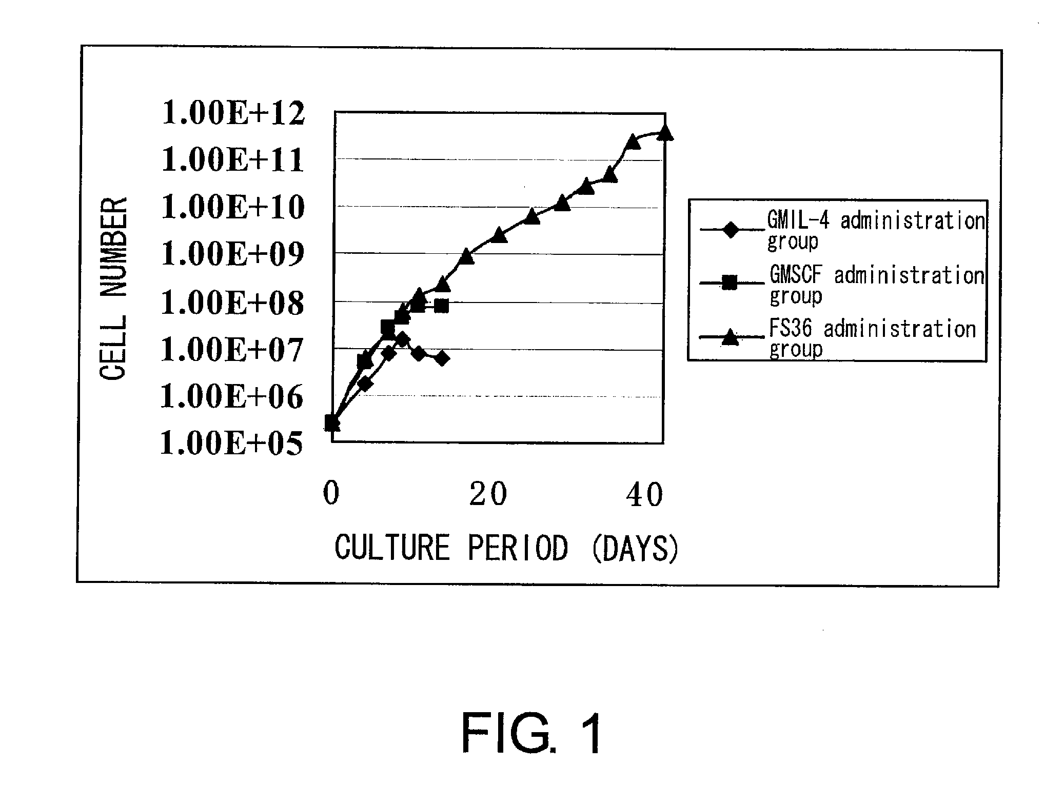
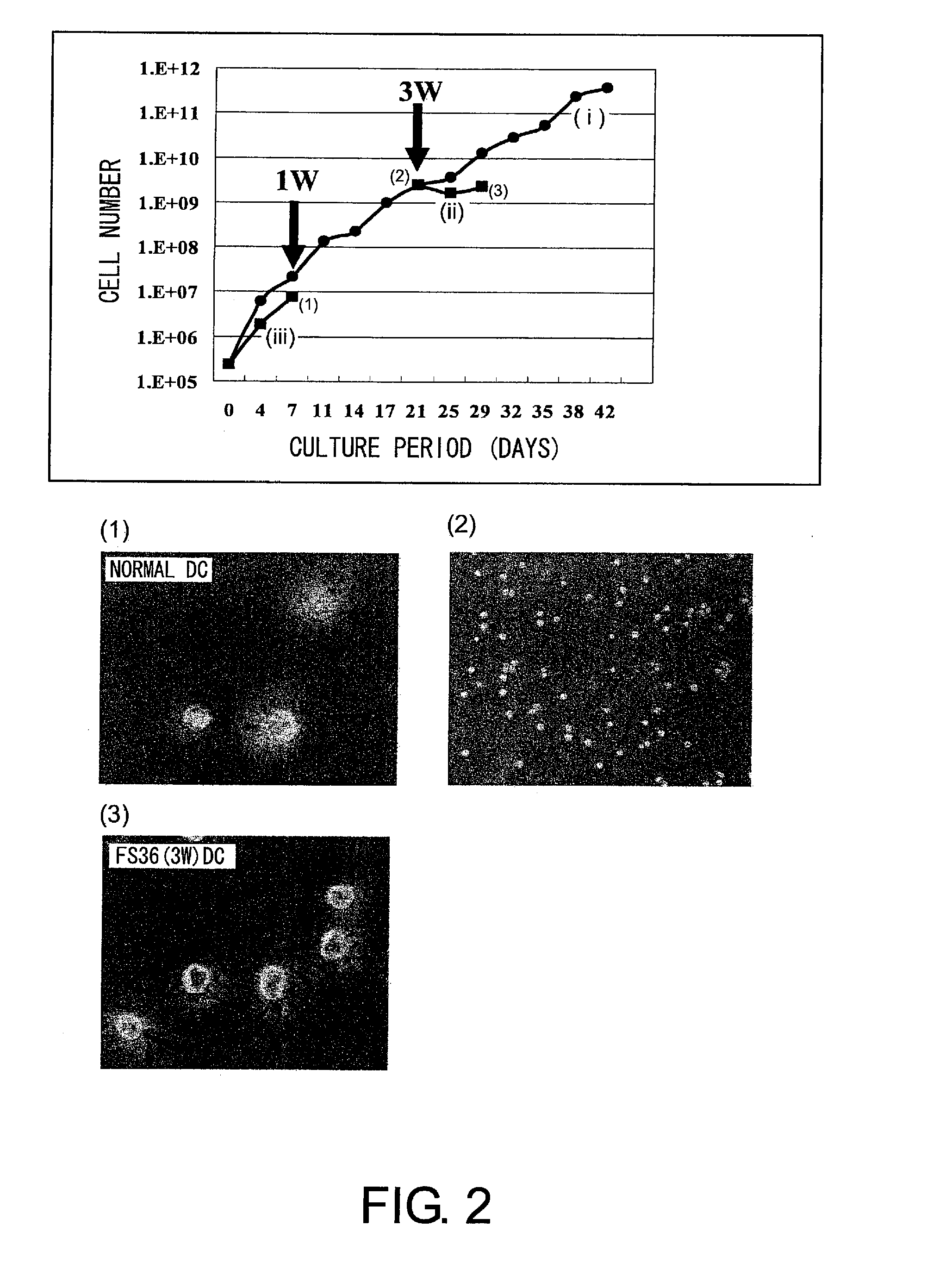
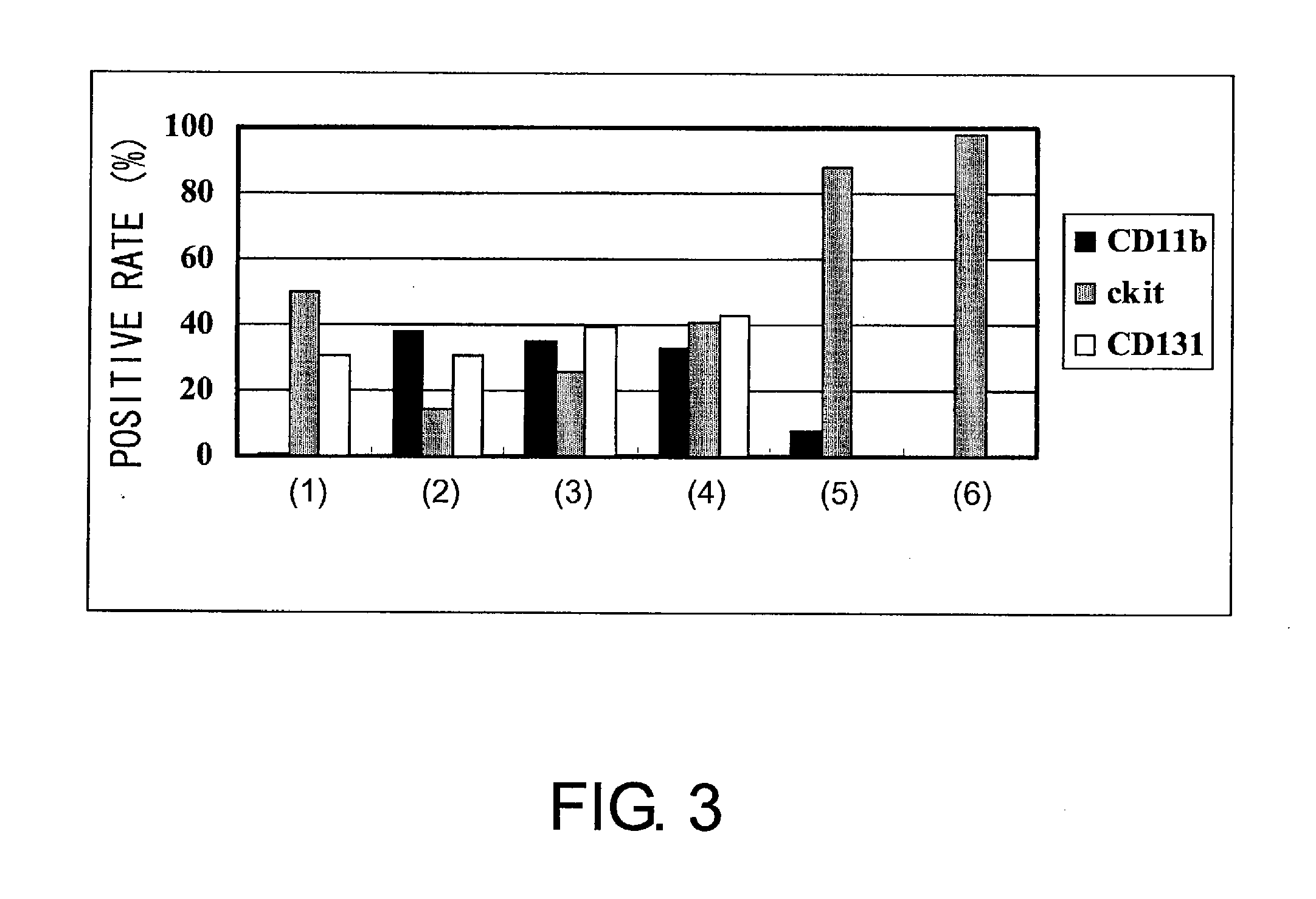
[0208]First, hematopoietic precursor cells were collected from the bone marrow of mouse (C3H) femur and tibia by negative selection (SpinSep mouse hematopoietic progenitor enrichment kit, StemCell technologies, Canada). The precursor cells were divided into three groups: FS36 administration group, GMIL-4 administration group, and GMSCF administration group. Then, the cells were cultured. The culture was started with 2.4×10⁵ cells. The cells were passaged every three or four days to have a concentration of 2×10⁶ cells / ml or lower, and the culture was continued for up to six weeks. Dendritic cell (DC) precursor cells were prepared in this process (FIG. 1). During this time, cells were counted to determine the growth rate. In addition, the differentiation ability of the above precursor cells was verified by FACS analysis after staining with anti-CD11b-FITC, anti-CD11c-PE, anti-c-kit-PE, ...
example 2
Differentiation of DC
[0213]DCs obtained from mouse hematopoietic precursor cells were infected with F gene-deficient Sendai virus (SeV / dF) at an moi of 50. Alternatively, LPS (1 μg / ml) was added to the DCs. Then, the cells were cultured for two days, and analyzed for the expression of DC surface markers with a flow cytometer using CD80-PerCP, CD86-PerCP, MHC classII-PerCP, and CD40-PerCP (FIGS. 11 and 12). The result showed that like normal DCs (FIG. 11(B)), when infected with Sendai virus or treated with LPS, DCs produced by one week of culture under the medium condition of the GMIL-4 administration group following three weeks of culture under the condition of the FS36 administration group expressed the co-stimulatory molecules CD80 and CD86, MHC Class II, and adhesion molecule (CD40) (FIG. 11(A)).
example 3
Expansion of DCs by GM-CSF and SCF
[0214]Human cord blood CD34+ cells (purchased from Cambrex) were expanded and differentiated by 35 days of culture under the condition of the GMSCF administration group or GMIL-4 administration group (1). During the culture period, the expression of c-kit, CD11c, and CD86 was analyzed using a flow cytometer every three to seven days of culture. When 1×10⁵ human cord blood CD34+ cells were cultured in a medium added with GM-CSF and SCF, CD11c+ cells grew gradually and 3.8×10⁹ cells were obtained after 35 days. Moreover, LPS was added on day 32, and FACS was carried out three days later using CD11c-PE and CD86-PE. The result showed that the expression of CD86 was enhanced by LPS (FIG. 16), similarly to the result described above in Example 2. Thus, both mouse and human CD11c+ cells can be expanded by using cytokine cocktails (FIGS. 13, 14, and 15).
PUM
| Property | Measurement | Unit |
|---|---|---|
| concentration | aaaaa | aaaaa |
| concentration | aaaaa | aaaaa |
| concentration | aaaaa | aaaaa |
Abstract
Description
Claims
Application Information
 - R&D
- Intellectual Property
- Life Sciences
- Materials
- Tech Scout
- Unparalleled Data Quality
- Higher Quality Content
- 60% Fewer Hallucinations
Browse by: Latest US Patents, China's latest patents, Technical Efficacy Thesaurus, Application Domain, Technology Topic, Popular Technical Reports.
© 2025 PatSnap. All rights reserved.Legal|Privacy policy|Modern Slavery Act Transparency Statement|Sitemap|About US| Contact US: help@patsnap.com


