
Method and System for Automatic Aorta Segmentation

Inactive Publication Date: 2010-09-23
SIEMENS HEALTHCARE GMBH
View PDF6 Cites 50 Cited by
  • Summary
  • Abstract
  • Description
  • Claims
  • Application Information

AI Technical Summary

Benefits of technology

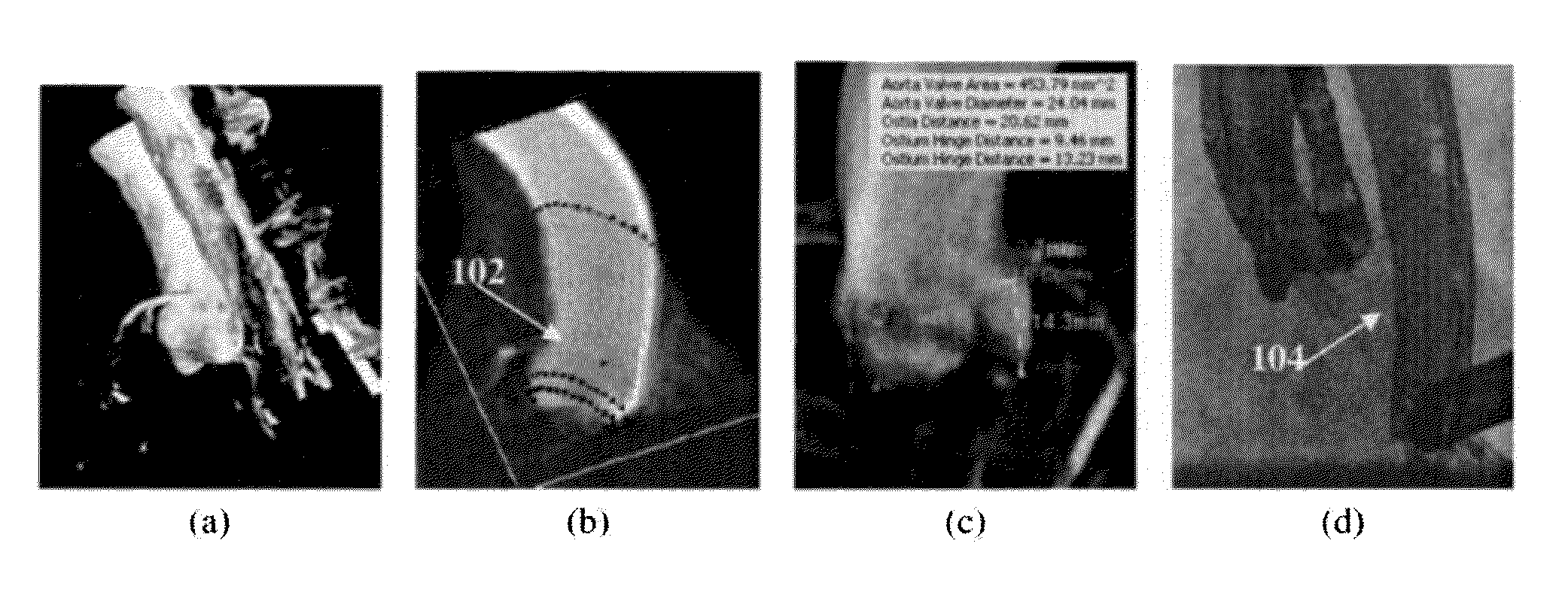
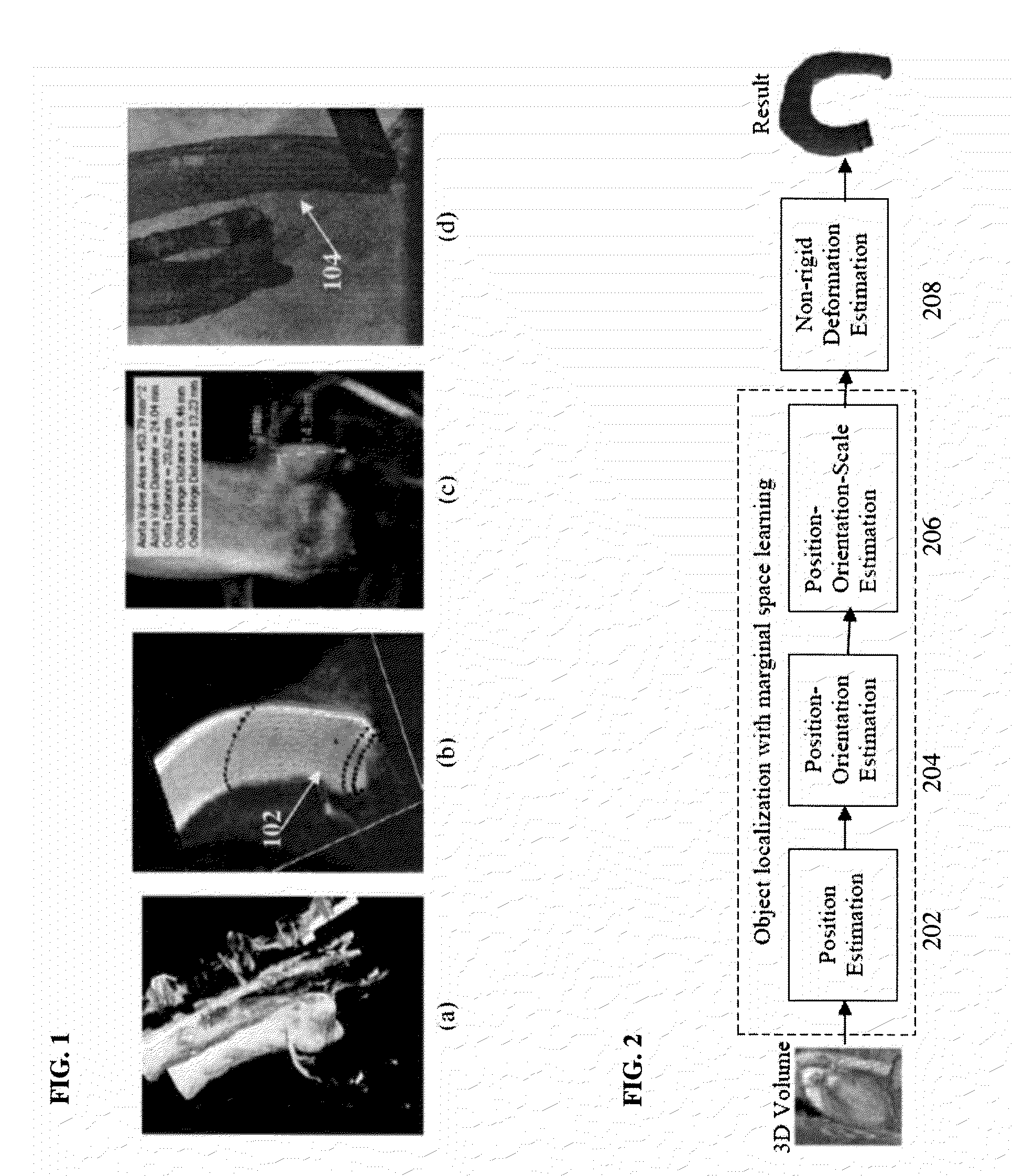
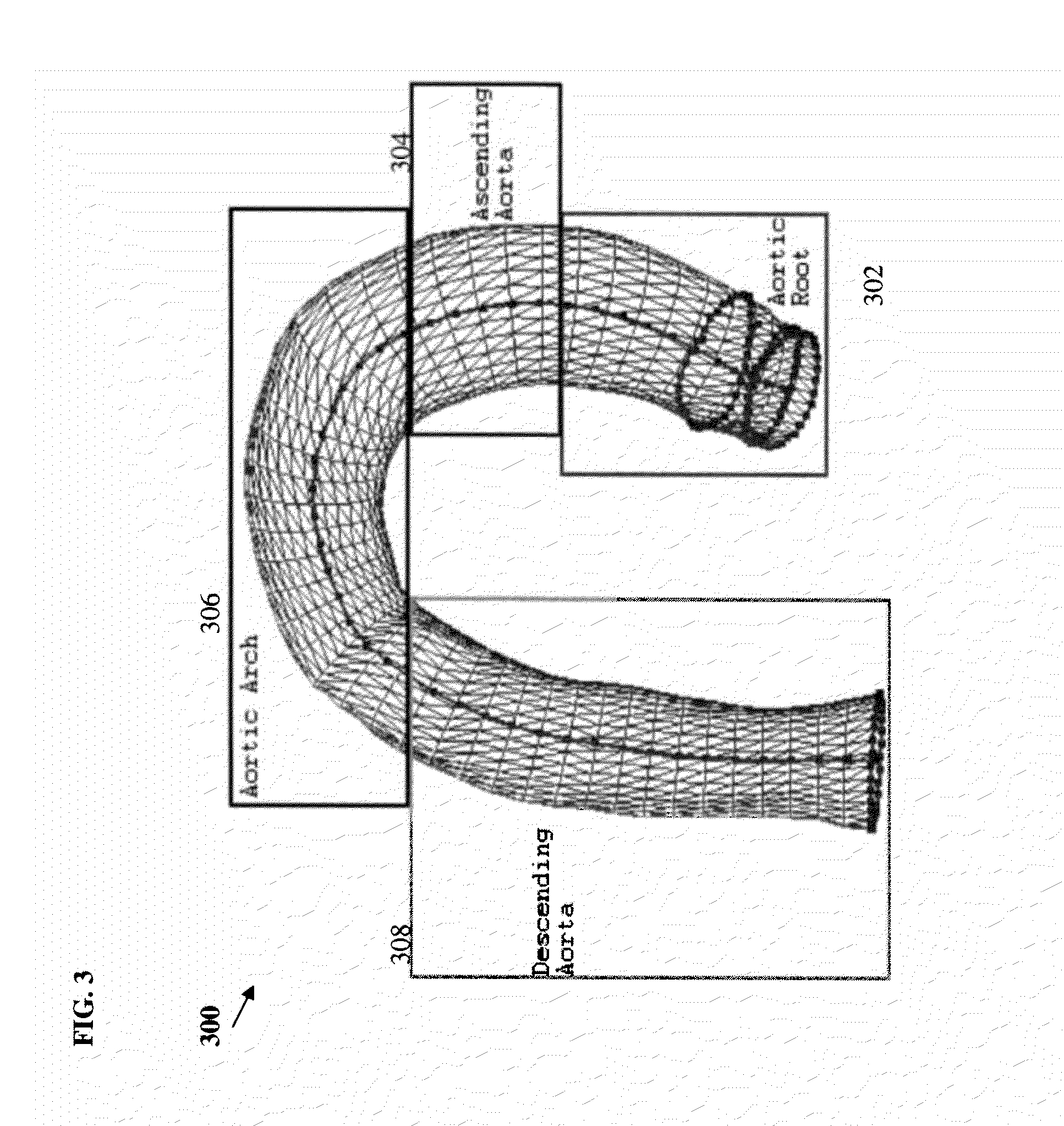
[0006]The present invention provides a method and system for automatic aorta segmentation that can be used to accurately segment the aorta in C-arm CT volumes. Embodiments of the present invention utilize a part-based approach in which the aorta is split into the aortic root, ascending aorta, aortic arch, and descending aorta. This approach is robust to structural variation and to variation of the use of contrast agents in C-arm CT volumes. The aorta segmentation results generated using embodiments of the present invention can be used in aortic valve implantation to provide 3D measurements of the aortic root, to provide the best angulation for a C-arm system for capturing 2D fluoroscopic images, and to overlay the segmented aorta onto the 2D fluoroscopic images to provide guidance during the implantation surgery.

Problems solved by technology

Although C-arm CT has many advantages over conventional CT with respect to artificial valve implantation surgery, automatic segmentation of the aorta in a C-arm CT volume is a challenging problem.
First, the image quality of a C-arm CT volume is typically not as good as a cardiac CT volume due to reconstruction artifacts and a large variation in the use of contrast agent.
For example, some C-arm CT volumes may have high contrast, while others have low contrast due to improper timing.
A simple intensity-based thresholding technique does not work for volumes having low contrast.
Accordingly, such a thresholding technique is not reliable for aorta segmentation in C-arm CT volumes.
Due to the variation in the aorta shape in various C-arm CT volumes, a statistical shape model cannot be applied directly for aorta segmentation.

Method used


Image

  • Method and System for Automatic Aorta Segmentation
  • Method and System for Automatic Aorta Segmentation
  • Method and System for Automatic Aorta Segmentation

Examples


Embodiment Construction

[0018]The present invention is directed to a method and system for automatic aorta segmentation in medical images, such as C-arm CT volumes. Embodiments of the present invention are described herein to give a visual understanding of the aorta segmentation method. A digital image is often composed of digital representations of one or more objects (or shapes). The digital representation of an object is often described herein in terms of identifying and manipulating the objects. Such manipulations are virtual manipulations accomplished in the memory or other circuitry / hardware of a computer system. Accordingly, it is to be understood that embodiments of the present invention may be performed within a computer system using data stored within the computer system.

[0019]Embodiments of the present invention provide a method for automatic aorta segmentation that can be used to accurately segment the aorta in C-arm CT volumes. This method is robust to structural variation and to variation of ...


PUM


Abstract

A method and system for aorta segmentation in a 3D volume, such as a C-arm CT volume is disclosed. The aortic root is detected in the 3D volume using marginal space learning (MSL) based segmentation. The aortic arch is detected in the 3D volume using MSL based segmentation. The ascending aorta is tracked from the aortic root to the aortic arch in the 3D volume, and the descending aorta is tracked from the aortic arch in the 3D volume.

Description

[0001]This application claims the benefit of U.S. Provisional Application No. 61 / 161,139, filed Mar. 18, 2009, the disclosure of which is herein incorporated by reference.BACKGROUND OF THE INVENTION[0002]The present invention relates to medical imaging of the heart, and more particularly, to automatic segmentation of the aorta in 3D medical images, such as C-arm CT volumes.[0003]Aortic valve disease is the most common valvular disease in developed countries, and has the second highest incidence among congenital valvular defects. Implantation of an artificial valve is often necessary to replace a damaged natural valve. Before surgery to replace a damaged valve, several important parameters of the aortic valve need to be extracted for surgery planning. For example, the diameter of the aortic valve annulus needs to be measured accurately in order to select an appropriately sized artificial valve. The distance from the coronary ostia to the sinutubular junction is also an important meas...

Claims


Application Information

Patent Timeline
IPC IPC(8): G06K9/00
CPCG06K9/4638G06K2209/051G06T2207/30101G06T2200/04G06T2207/10081G06T7/0083G06T7/12G06V10/457G06V2201/031
Inventor ZHENG, YEFENGGEORGESCU, BOGDANJOHN, MATTHIASBOESE, JANCOMANICIU, DORIN
Owner SIEMENS HEALTHCARE GMBH
Learn More