Eureka AIR delivers breakthrough ideas for toughest innovation challenges, trusted by R&D personnel around the world.

Method for detecting DNA methylation in cancer cells

a cancer cell and methylation technology, applied in the field of cancer detection methods, can solve the problems of difficult to determine if patients are in true remission, the approach has not been widely adopted in clinical setting, and the dna destruction of bisulfite is not easy to d

Inactive Publication Date: 2010-09-30
UNIVERSITY OF MISSOURI
View PDF4 Cites 7 Cited by
  • Summary
  • Abstract
  • Description
  • Claims
  • Application Information

AI Technical Summary

Benefits of technology

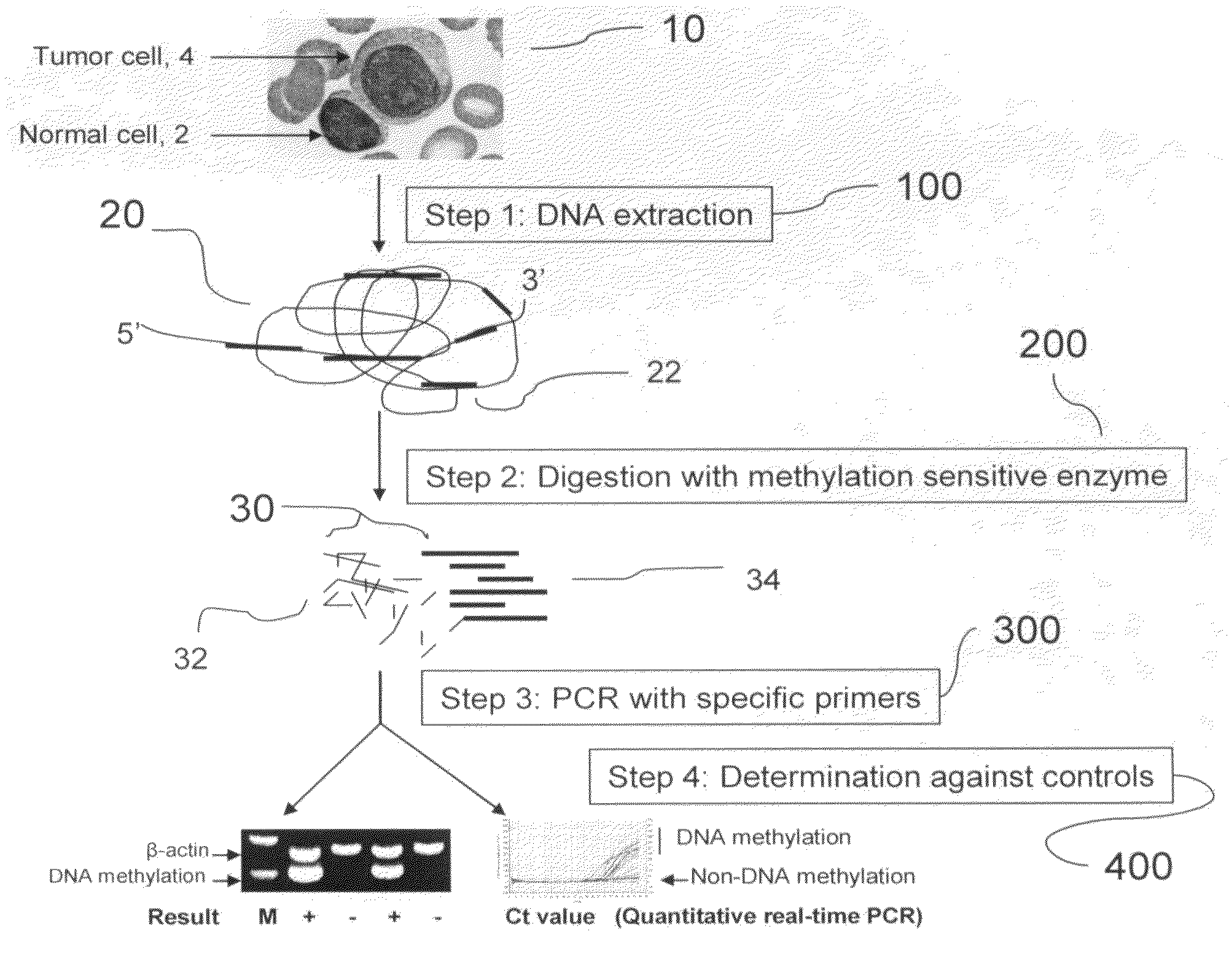
[0016]According to one embodiment of the inventive method, the DNA sample may be collected from a patient's blood, bone marrow, tissue, or other specimens; the methylation sensitive enzyme may be selected from Aci I, Hap II, HinP1 I, BstU I, Hha I, Tai I, or any combination thereof; and the primers may be selected from DLC-1 primers, CDH1 primers, PCDHGA12 primers, or any combination thereof. The preferred internal control for PCR amplification is β-actin primers. The positive control may be any known tumor cell line DNA or Sss I methyltransferase-treated normal human DNA. The negative control may be any normal human blood or bone marrow cell DNA.
[0017]According to another embodiment of the invention, the aforesa...

Problems solved by technology

However, bisulfite destroys the majority of DNA during the treatment.
However, this approach has not been widely adopted at clinical setting because of a high degree of false positive signal resulted by incomplete enzyme digestion.
After induction chemotherapy, however, to determine if the patients are in true remission is problematic.
If these residual leukemic cells are left with no further treatment, most patients will relapse.
Although these methods have high sensitivity (10−4 to 10−5) and specificity, they are unable to identify the residual leukemic cells that lack initial specific detectible chromosomal translocations or with ongoing somatic mutations resulting in clonal evolution or antigen shifting.
In addition, the technical complexity, poor reproducibility at higher sensitivity, and high cost are the major obstacles for the routine clinical application.

Method used


Image

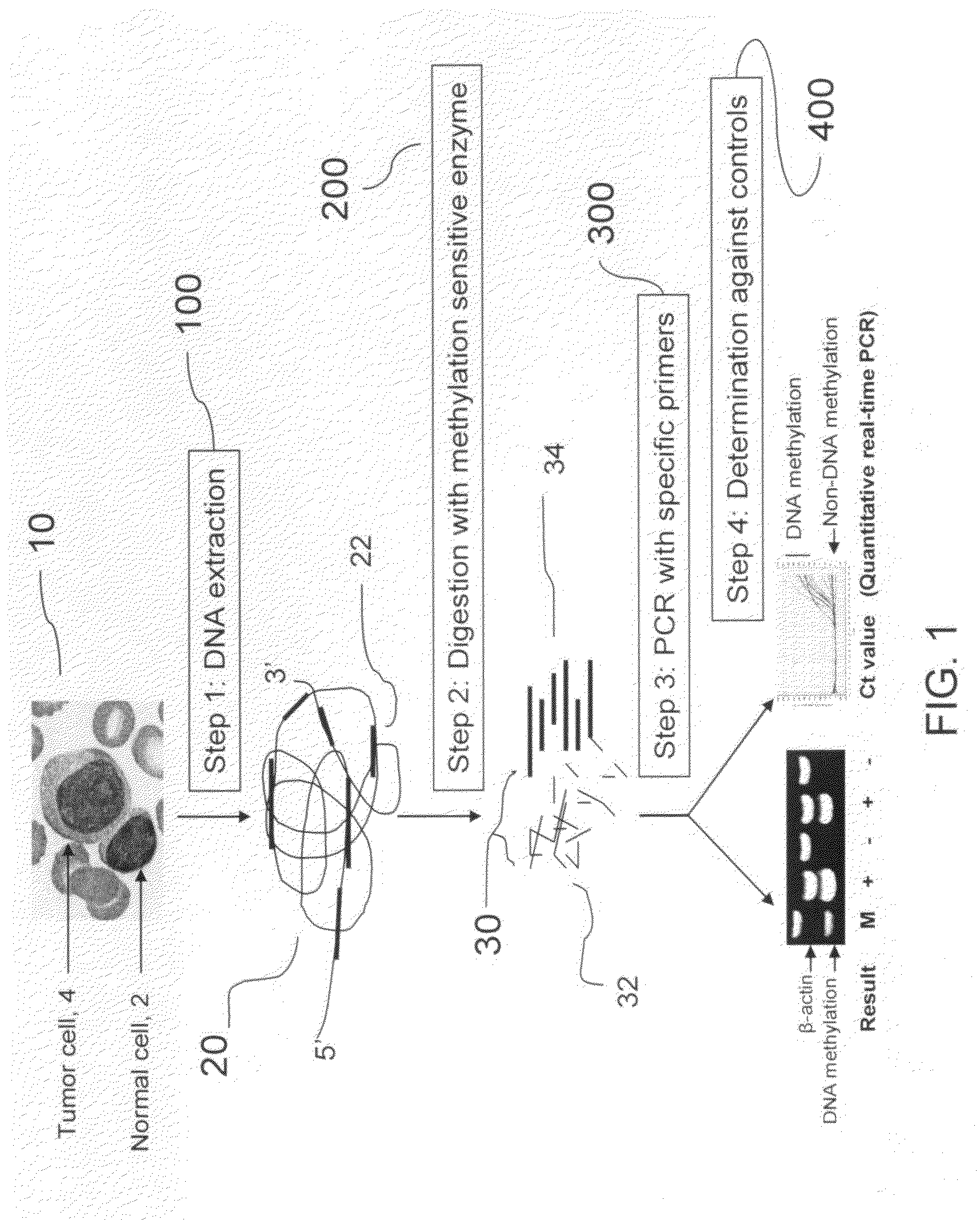
  • Method for detecting DNA methylation in cancer cells
  • Method for detecting DNA methylation in cancer cells
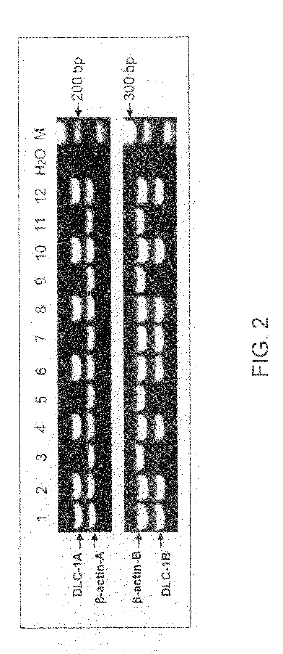
  • Method for detecting DNA methylation in cancer cells

Examples


Embodiment Construction

[0029]Unless otherwise defined, all technical and scientific terms used herein have the same meaning as commonly understood by one of ordinary skill in the art to which this invention belongs. All publications, patent applications, patents, and other references mentioned herein are incorporated by reference in their entirety.

[0030]The present invention provides a method for determining DNA methylation patterns at a cytosine residue of a CpG sequence by enzyme digestion of a sample of genomic DNA with one or multiple pre-selected methylation sensitive restriction enzymes followed by PCR amplification with one or multiple pre-selected primers. The invention teaches that there is fundamental difference between malignant and normal cells in their methylomes. When a genomic DNA sample with malignant and normal cells is subjected for methylation sensitive enzyme restriction, their patterns of digestion will be different. Specific hypermethylation regions in malignant cells are resistant t...


PUM

PropertyMeasurementUnit
Fractionaaaaaaaaaa
Timeaaaaaaaaaa
Biological propertiesaaaaaaaaaa

Abstract

The present invention provides a detecting method of detecting malignant cells in a patient's specimen or a biological sample. Specifically, the inventive method includes the steps of extracting a genomic DNA, digesting said genomic DNA with one or multiple methylation sensitive restriction enzymes, and amplifying by PCR with one or multiple selected primers. The PCR can be performed in a conventional or a real-time platform. The inventive method can detect leukemia cells in 90% ALL patients at a sensitivity of up to 10−6. The inventive method also provides broad clinical applications in cancer (including hematopoietic and solid tumors) screening and risk assessment, early detection and diagnosis confirmation, and therapeutic monitoring, minimal residual disease detection and prognostic prediction.

Description

CROSS-REFERENCE TO RELATED APPLICATIONS[0001]This application claims the benefit of U.S. Provisional Application Ser. No. 61 / 072,064, entitled “Method for Detecting DNA Methylation in Cancer Cells,” to Wang, et al., filed on Mar. 27, 2008.BACKGROUND OF INVENTION[0002]1. Field of the Invention[0003]The present invention relates to a cancer detection method based upon DNA methylation differences at specific CpG sites. More specifically, the present invention relates to a diagnostic method for detecting cancer cells in cancer patients, especially for acute lymphoblastic leukemia.[0004]2. Background Art[0005]Methods in Detecting Aberrant DNA Methylation[0006]In the past decade, the studies in the cancer epigenetic area have identified hundreds of aberrant DNA methylation loci in virtually all types of cancers including hematopoietc tumors. DNA hypermethylation usually occurs in CG rich promoter region and / or exon 1 region (CpG island) of a gene and is tumor specific and inheritable. If ...

Claims


Application Information

Patent Timeline
IPC IPC(8): C12Q1/68
CPCC12Q1/6883C12Q2600/16C12Q2600/154
Inventor WANG, MICHAEL XIASHI, HUIDONGNALLURI, SRILATHA
Owner UNIVERSITY OF MISSOURI
Why Eureka
Eureka Blog
Learn More