Orthodontic treatment monitoring based on reduced images

Active Publication Date: 2010-10-14
3M INNOVATIVE PROPERTIES CO
View PDF25 Cites 84 Cited by
  • Summary
  • Abstract
  • Description
  • Claims
  • Application Information

AI Technical Summary

Benefits of technology

[0007]Due to the limitations in conventional orthodontic treatment monitoring methods described above, the process of providing precise relative positions of teeth during treatment is often time consuming and inefficient. The present invention provides methods to conveniently and efficiently monitor and evaluate the positions of a patient's teeth during the course of orthodontic treatment using digital information. In an exemplary embodiment, an initial set of digital data representing a patient's dental structure is provided by an X-ray radiograph and/or intraoral scan. Then during the course of treatment, a reduce

Problems solved by technology

Due to the limitations in conventional orthodontic treatment monitoring methods described above, the process of providing precise relative positions of teeth during treatment is often time consuming and inefficient.
First, by leveraging a full set of dental data already acquired at the outset of treatment, only small por

Method used


Image

  • Orthodontic treatment monitoring based on reduced images
  • Orthodontic treatment monitoring based on reduced images
  • Orthodontic treatment monitoring based on reduced images

Examples


example

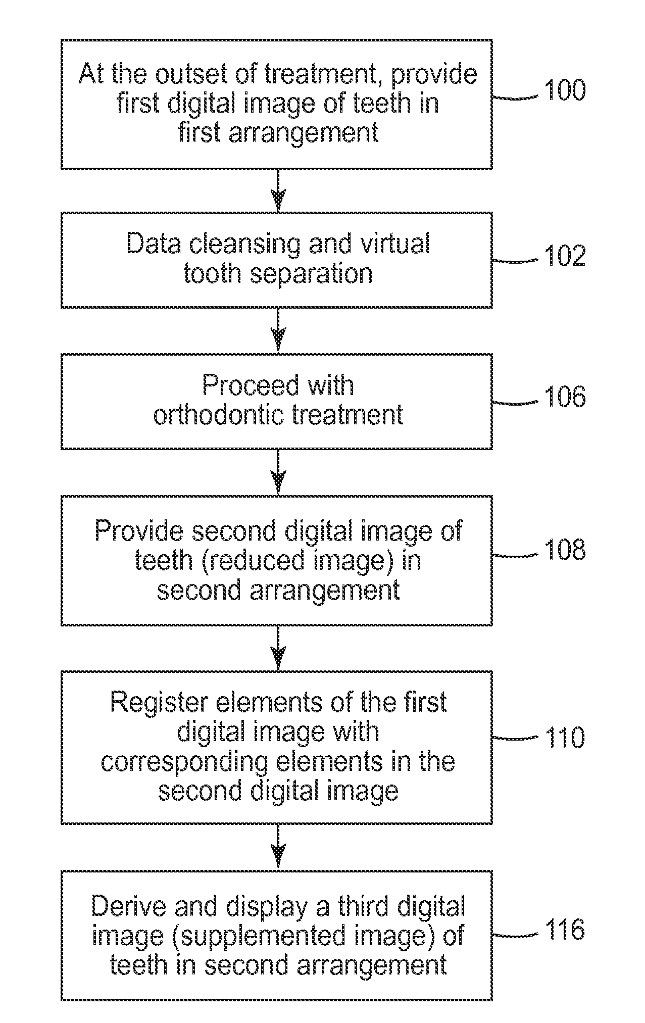
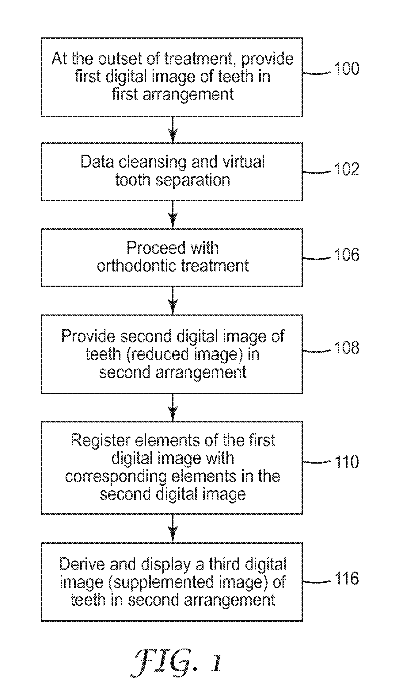
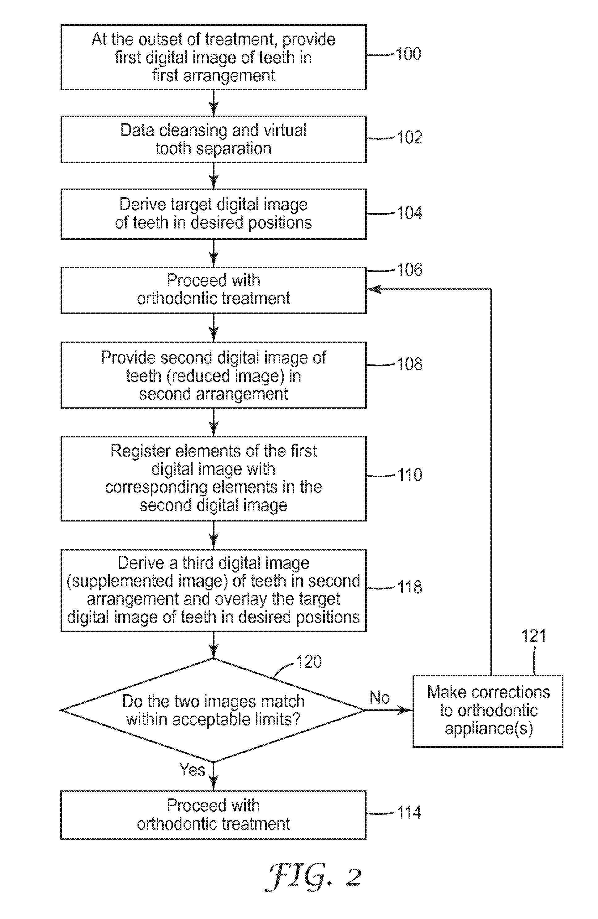
[0084]Aspects of the methods above are further demonstrated by the following example. A physical dental model of the upper and lower jaws, made from orthodontic stone, was provided by a local orthodontic office. For exemplary purposes, this dental model was used to simulate a patient's dental structure in a given dental arrangement. To prepare the first digital image, a polyvinylsiloxane impression was first taken using POSITION PENTAQUICK brand impression material available from 3M ESPE (St. Paul, Minn.). After allowing the impression to set for 90 seconds, it was removed from the dental model and used to cast a duplicate positive model from F82-302 epoxy resin from United Resin Corporation (Royal Oak, Mich.). To impart opacity to the epoxy model, it was coated with a fine layer of SPOTCHECK brand SKD-S2 Developer white talc powder from Magnaflux (Genview, Ill.). The coated model was then scanned using a SURVEYOR brand OM-3R narrow spectrum visible light laser scanner from Laser De...


PUM


Abstract

The present invention provides digitally driven methods to conveniently and efficiently monitor and evaluate the positions of a patient's teeth during the course of orthodontic treatment. In an exemplary embodiment, an initial set of 3D digital data representing a patient's dental structure is provided by means of an X-ray radiograph and/or an intraoral scan. Then during the course of treatment, a reduced image representing part of the dental structure may be quickly provided using a bite plate or a low resolution intraoral scan. A three-dimensional (3D) image of the full dental structure is subsequently rendered or ‘reconstructed’ by registering elements of the initial image with corresponding elements of the reduced image. By reconstructing the dental structure from the reduced image, the treating professional can provide a precise, manipulable 3D image of the patient's complete dental structure for diagnostic and treatment planning purposes. Other aspects include methods of using these data to compare actual and target teeth positions to evaluate the progress of treatment and suggest revisions to the specification of an orthodontic appliance.

Description

FIELD OF THE INVENTION [0001]The present invention is generally in the field of orthodontic treatment. More particularly, the present invention is directed to methods of orthodontic treatment monitoring and planning.DESCRIPTION OF THE RELATED ART [0002]Orthodontics is the area and specialty of dentistry associated with the supervision, guidance, and correction of teeth positions in the mouth. This is achieved by the application and / or re-direction of forces to teeth to bring them into proper positions relative to each other as well as facial bones such as the jawbone.[0003]In one common type of treatment, tiny slotted appliances called brackets are affixed to a patient's teeth and a resilient archwire is placed in the slot of each bracket. The ends of the archwire are provided in appliances called buccal tubes, which are affixed to the patient's molar teeth. The archwire is deflected when placed in the mouth, but subsequently imparts continuous forces to urge the teeth to proper loc...

Claims


Application Information

Patent Timeline
IPC IPC(8): G06K9/62
CPCA61C7/002A61C7/00
Inventor CINADER, JR., DAVID K.
Owner 3M INNOVATIVE PROPERTIES CO
Why Eureka
Try Eureka