Systems and methods for imaging a blood vessel using temperature sensitive magnetic resonance imaging

a technology of magnetic resonance imaging and blood vessel, applied in the field of system and method for imaging a blood vessel using temperature sensitive magnetic resonance imaging, can solve the problems of inability to easily repeat both conventional x-ray angiography and ct angiography, adverse effects of iodinated contrast material injection,

Inactive Publication Date: 2010-10-21
THE TRUSTEES OF COLUMBIA UNIV IN THE CITY OF NEW YORK
View PDF10 Cites 9 Cited by
  • Summary
  • Abstract
  • Description
  • Claims
  • Application Information

AI Technical Summary

Problems solved by technology

Conventional x-ray angiography is an invasive procedure and the injection of iodinated contrast material can be associated with adverse reactions including severe allergic reactions and anaphylaxis.
However, CT angiography still requires the use of x-rays.
In addition, because they require iodinated contrast agents, both conventional x-ray angiography and CT angiography cannot be readily repeated.
However, ultrasound angiography has limited spatial resolution, limited depth of penetration into the body and does not readily allow multiplanar reconstruction.
In addition, ultrasound angiography cannot visualize the cerebral vasculature,
Although these contrast agents are safer than iodinated agents, they still carry

Method used


Image

  • Systems and methods for imaging a blood vessel using temperature sensitive magnetic resonance imaging
  • Systems and methods for imaging a blood vessel using temperature sensitive magnetic resonance imaging
  • Systems and methods for imaging a blood vessel using temperature sensitive magnetic resonance imaging

Examples


Embodiment Construction

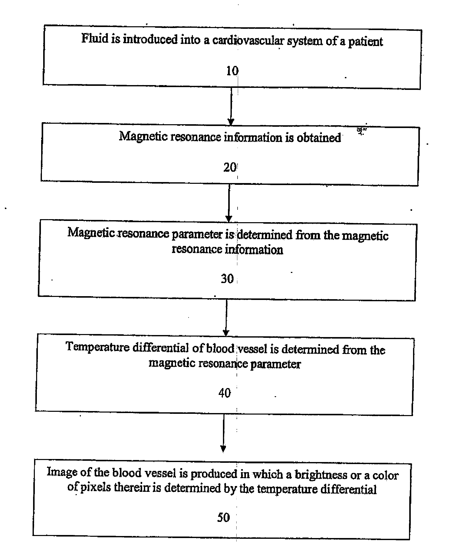
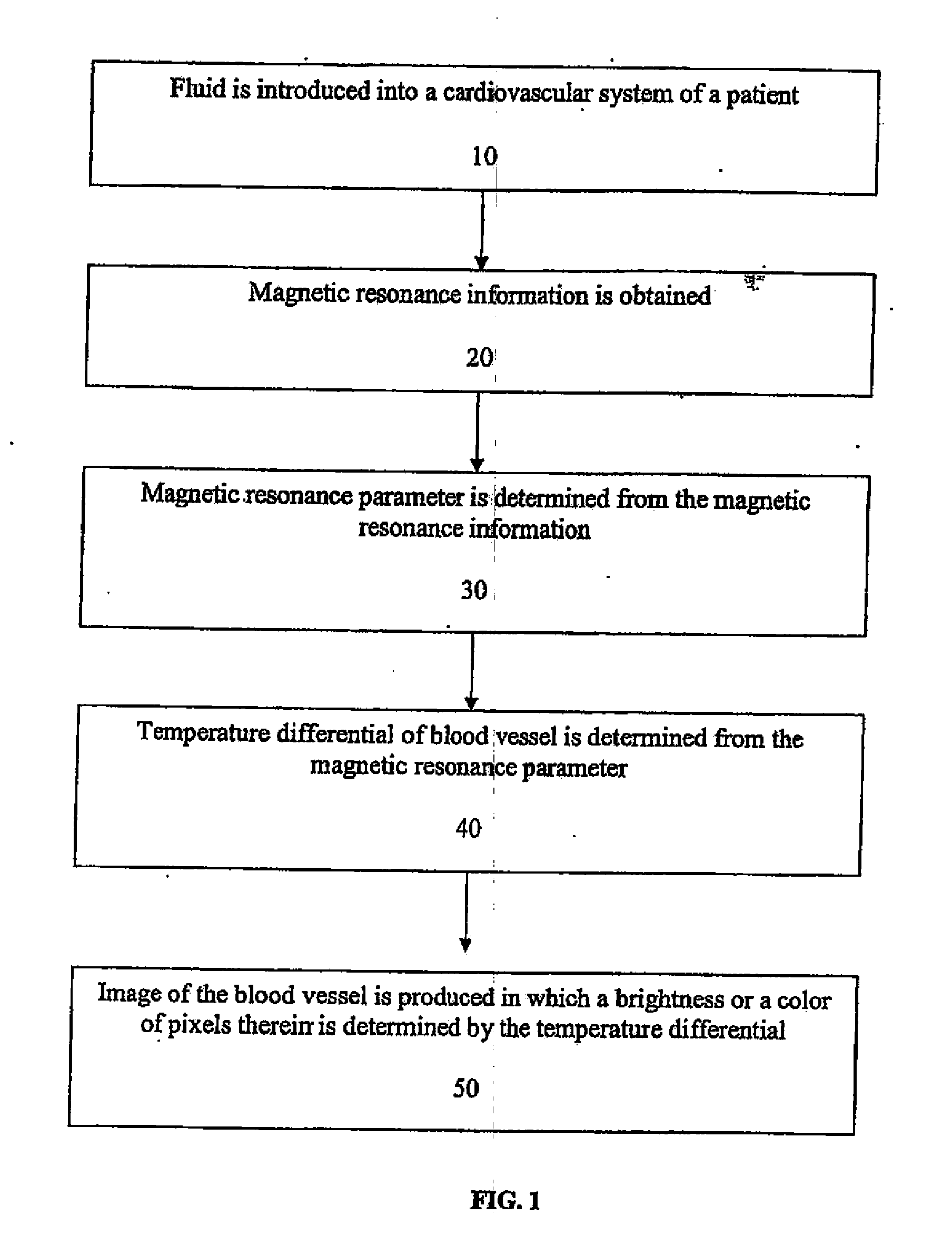
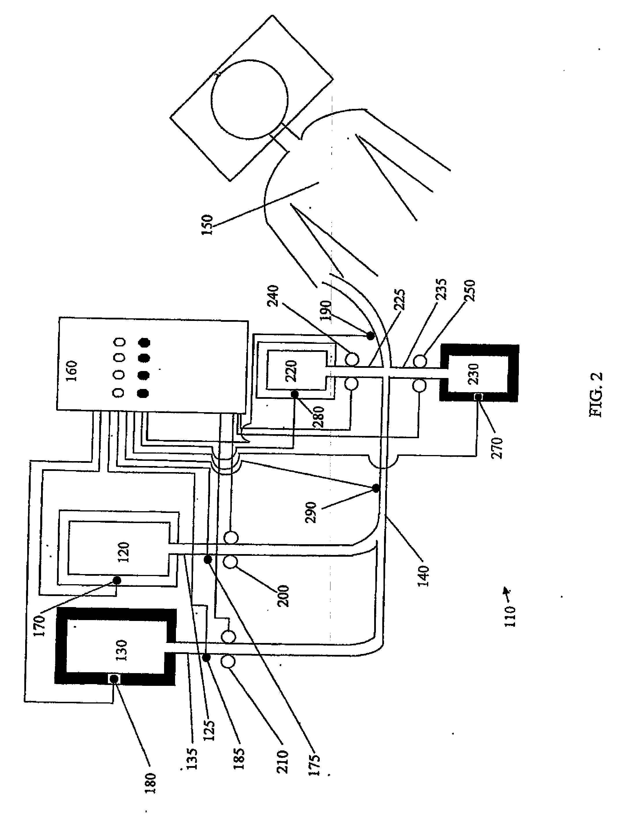
[0014]In an embodiment, the present invention provides a method for producing an image of a blood vessel of a patient based on a temperature differential of flowing blood within the vessel determined from information obtained by MRI. Specifically, referring to FIG. 1, a method for producing an image of a blood vessel comprises introducing a fluid into a cardiovascular system of a patient (10) and then obtaining magnetic resonance information from the blood vessel of the patient (20). A magnetic resonance parameter is determined using the magnetic resonance information (30) and a temperature differential in the blood vessel is determined using the magnetic resonance parameter (40). Based on the temperature differential, an image of the blood vessel is produced in which a brightness or a color of pixels therein is determined by the temperature differential (50).

[0015]The blood vessel can be a part of the vasculature of a patient including an artery, vein, capillary or combination ther...


PUM


Abstract

A method for producing an image of a blood vessel in a patient utilizing temperature sensitive MRI measurement. The method includes introducing a fluid in a blood vessel, obtaining magnetic resonance information from the blood vessel, and determining a magnetic resonance parameter using the magnetic resonance information. The method further includes using the magnetic resonance parameter to determine a temperature differential in the blood vessel and producing an image of the blood vessel based on the temperature differential. Systems for producing an image of a blood vessel in a patient using temperature sensitive MRI measurements are also provided.

Description

CROSS-REFERENCE TO RELATED APPLICATIONS[0001]The present application claims the benefit of and priority to International Patent Application No. PCT / US07 / 02032, filed 22 Jan. 2007, which claims the benefit of and priority to U.S. Provisional Patent Application No. 60 / 761,773, filed 25 Jan. 2006, both of which are expressly incorporated herein in their entireties by reference thereto.[0002]The present application is related to co-pending applications “Systems and Methods for Determining a Cardiovascular Parameter Using Temperature Sensitive Magnetic Resonance Imaging,” filed herewith and “Systems and Methods for Determining Metabolic Rate Using Temperature Sensitive Magnetic Resonance. Imaging,” filed herewith. Both applications are incorporated by reference herein.FIELD OF THE INVENTION[0003]The present invention relates to methods and systems for producing an image of a blood vessel based on a temperature differential determined from information obtained by magnetic resonance imagin...

Claims


Application Information

Patent Timeline
IPC IPC(8): A61B5/055
CPCA61B5/02007A61B5/411A61B5/055A61B5/028
Inventor PILE-SPELLMAN, JOHNLIN, ERWIN
Owner THE TRUSTEES OF COLUMBIA UNIV IN THE CITY OF NEW YORK
Why Eureka
Try Eureka