Methods and Apparatus for Detecting and Mapping Tissue Interfaces

a technology of tissue interfaces and apparatus, applied in the field of tissue mapping, can solve the problems of reducing accuracy and usefulness for practitioners, information is presently not readily available to practitioners, etc., and achieves the effect of improving accuracy and facilitating the measurement of tissue thickness

Inactive Publication Date: 2011-02-03
ST JUDE MEDICAL ATRIAL FIBRILLATION DIV
View PDF21 Cites 62 Cited by
  • Summary
  • Abstract
  • Description
  • Claims
  • Application Information

AI Technical Summary

Benefits of technology

[0021]An advantage of the present invention is that it facilitates measuring tissue thicknesses with increased accuracy.
[0022]Another advantage of the present invention is that it associates measured tissue thicknesses with particular spatial coordinates. The resultant plurality of data points may be used to generate maps and / or graphical representations of tissue thickness.
[0023]Still another advantage of the present invention is that acoustic echo measurements are often more accurate than measurements automatically or manually derived from presurgical images (e.g., CATSCAN or MRI images).

Problems solved by technology

Unfortunately, such information is presently not readily available to practitioners.
While it is known to estimate thickness information from dual-axis fluoroscopic images or based upon the practitioner's histological experience, these methods are inherently subjective and therefore of reduced accuracy and usefulness to practitioners.

Method used


Image

  • Methods and Apparatus for Detecting and Mapping Tissue Interfaces
  • Methods and Apparatus for Detecting and Mapping Tissue Interfaces
  • Methods and Apparatus for Detecting and Mapping Tissue Interfaces

Examples


Embodiment Construction

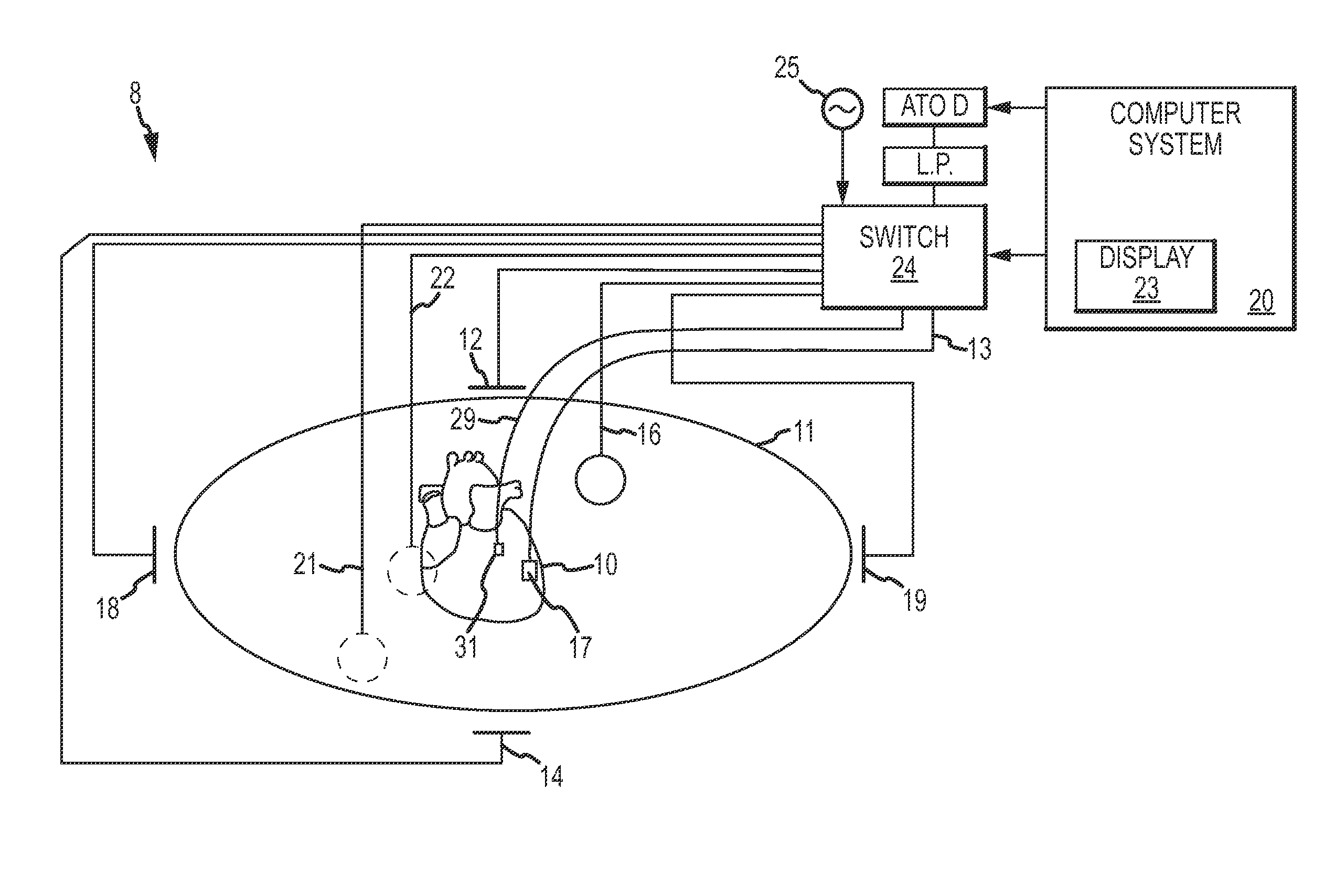
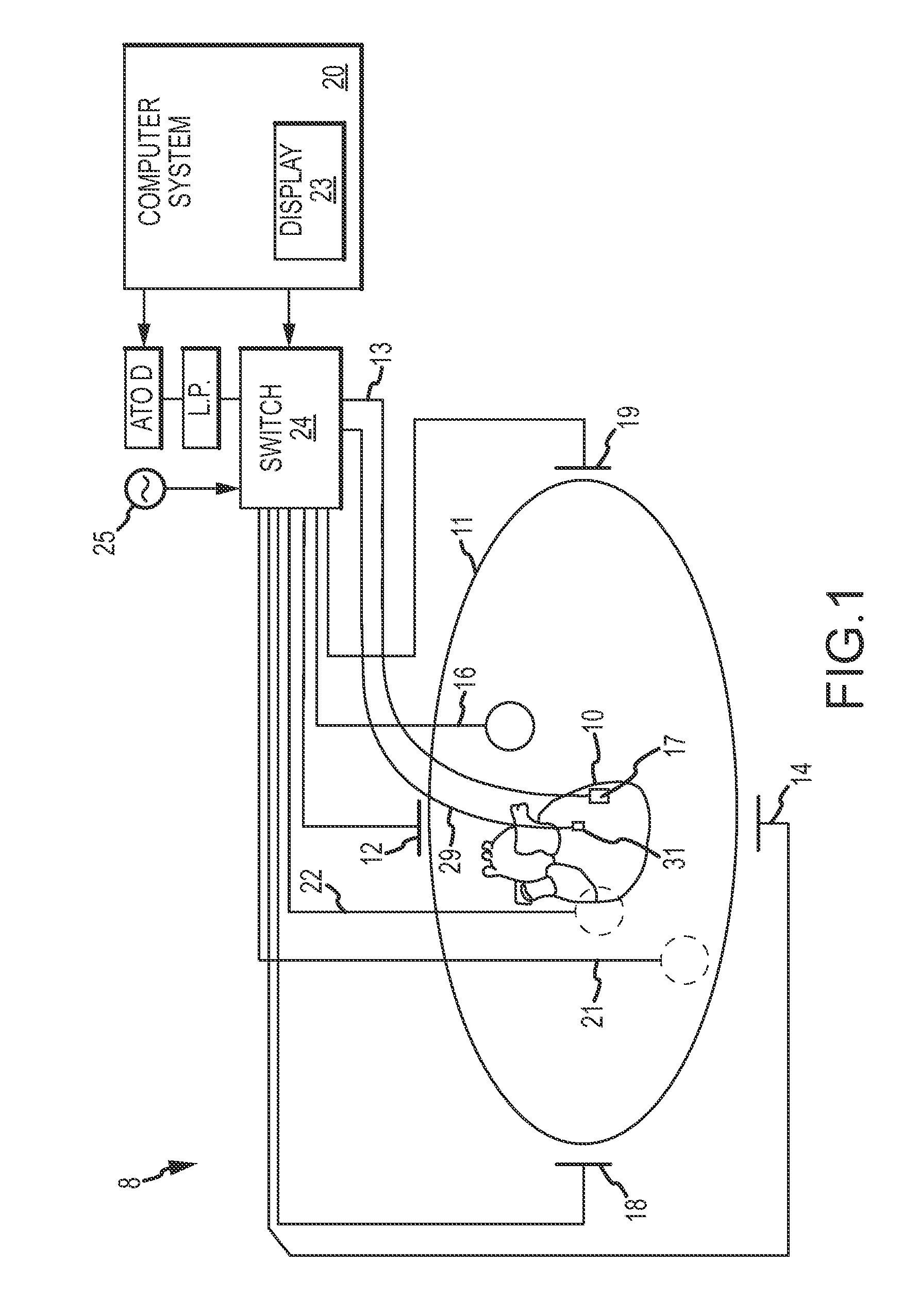
[0031]The present invention provides a method and system for locating tissue interfaces and locations where diverse tissue meets other tissue (e.g., tissue having different density or the like) or where body fluids are the dominant adjoining anatomical “structure,” for example in order to map tissue or fluid volumes and / or to measure tissue thicknesses. For purposes of illustration, the invention will be described in detail in the context of measuring and mapping cardiac tissue thickness. It is contemplated, however, that the present invention may be practiced to good advantage in other contexts, such as the detection of potential aneurysms via the detection of thinned cardiac tissue or the detection of relatively thick chamber tissue indicating a potentially deleterious progression of heart failure or other circulatory disease. The present invention may also be practiced to good advantage to gather data relating to variations in tissue thickness over time (e.g., thickness vs. heart...


PUM


Abstract

A device for measuring a spatial location of a tissue surface, such as the interface between different types of tissues or between tissue and body fluids, generally includes an elongate catheter body having a distal end portion, a plurality of localization elements carried by the distal end portion, and at least one pulse-echo acoustic element carried by the distal end portion. The localization elements allow the catheter to be localized (e.g., position and / or orientation) within a localization field, while the acoustic element allows for the detection of tissue surfaces where incoming acoustic energy will reflect towards the acoustic element. A suitable controller can determine the location of the detected tissue surface from the localization of the distal end portion of the catheter body. Tissue thicknesses can be derived from the detected locations of multiple (e.g., near and far) tissue surfaces. Maps and models of tissue thickness can also be generated.

Description

BACKGROUND OF THE INVENTION[0001]a. Field of the Invention[0002]The instant disclosure relates to tissue mapping. In particular, this disclosure relates to apparatus and methods for detecting, locating, and mapping tissue interfaces, for example to generate maps and models of tissue thickness and optionally indices of patient condition.[0003]b. Background Art[0004]It is well known to utilize images and / or geometric models of a patient's heart chamber(s) in conjunction with cardiac diagnostic or therapeutic procedures, such as ablation procedures to treat atrial fibrillation and other cardiac rhythm disorders. It is also known to provide these images and / or geometric models with electrophysiological data, such as maps of electrical potential, overlaid thereon.[0005]Information concerning the thickness of the tissue being mapped and / or treated may also be useful to a practitioner. Unfortunately, such information is presently not readily available to practitioners. While it is known to...

Claims


Application Information

Patent Timeline
Patent Type & Authority Applications(United States)
IPC IPC(8): A61B8/14
CPCA61B6/503A61B6/541A61B8/0883A61B8/12A61B8/4245A61B8/543A61B8/445A61B2017/00106A61B2018/00357A61B2018/00577A61B2019/5251A61N7/022A61B18/1492A61B2034/2051
Inventor SHAQUER, CEMSLIWA, JOHN W.MA, ZHENYIDANDO, JEREMY D.
Owner ST JUDE MEDICAL ATRIAL FIBRILLATION DIV
Why Eureka
Try Eureka