

Malignancy-risk signature from histologically normal breast tissue
a breast tissue and risk signature technology, applied in the field of cancer screening and therapy, can solve the problems of considerable cosmetic defects in the residual breast, and the risk of cancer not completely eliminated
- Summary
- Abstract
- Description
- Claims
- Application Information
AI Technical Summary
Benefits of technology
Problems solved by technology
Method used
Image

Examples
example 1
Materials and Methods
[0146]Tissues were collected in accordance with the protocols approved by the Institutional Review Board of the University of South Florida, and stored in the tissue bank of Moffitt Cancer Center. Breast tissues from patients that underwent mastectomy at various stages of breast carcinoma were collected and frozen in liquid nitrogen. The tissues were embedded in Tissue-Tek® O.C.T., 5-μm sections cut and mounted on Mercedes Platinum StarFrost™ Adhesive slides. The slides were stained using a standard H&E protocol, and tissue boundaries marked. Using the marked slide as a “map”, tissues were microdissected. Adipose tissues were trimmed away; the tumor and “normal” tissues were separated and stored in liquid nitrogen.
[0147]Histological examination of all tissue sections and microdissection of samples were conducted by pathologist to ensure consistency in the clinical diagnoses. From a large invasive breast cancer database, a set of 42 hist...
example 2
IDC Gene Signature
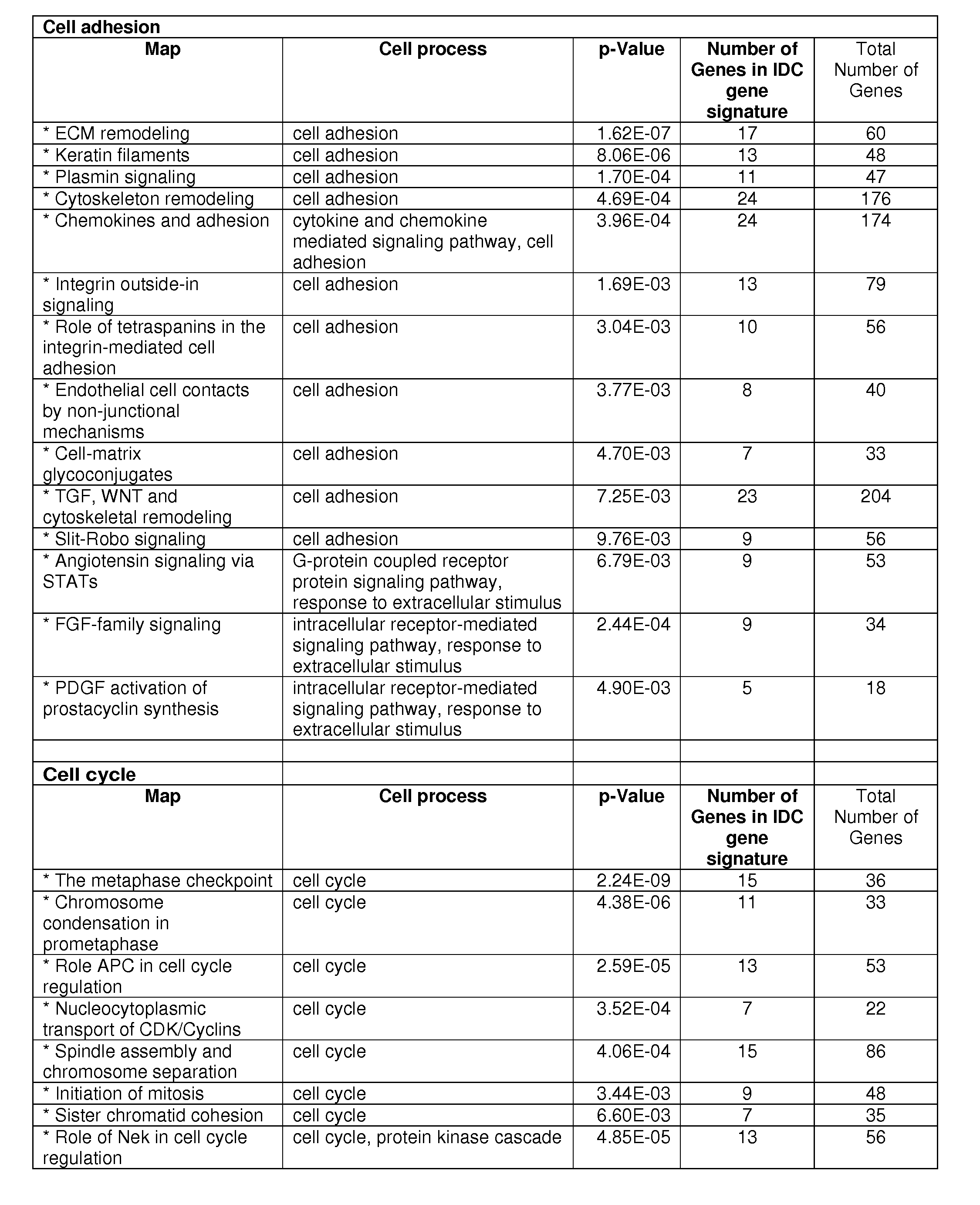
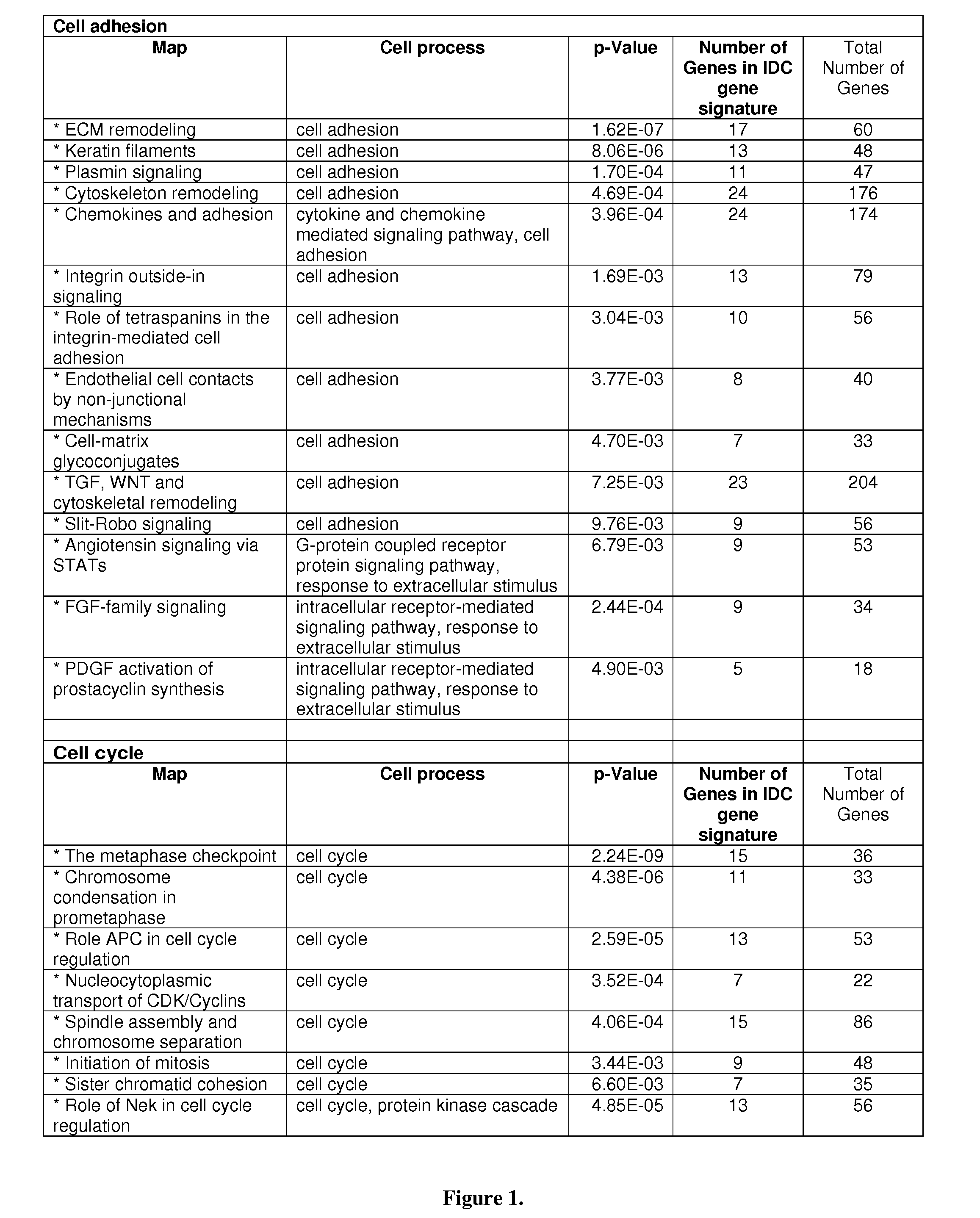
[0151]An IDC gene signature (1,554 probe sets: 1038 unique genes) was first developed from a set of 42 IDC and 143 normal breast tissues. This analysis was done using Statistical Analysis of Microarray7 and based on a cutoff of false discovery rate (FDR) 2. Pathway analysis revealed two predominant cellular processes: cell cycle and cell adhesion, as seen in FIG. 1. There were 10 cell adhesion pathways and 7 cell cycle pathways with a significant p-value <0.01. A majority of the genes were down-regulated in the cell adhesion, but up-regulated in the cell cycle.
example 3
Outlier Breast Tissues
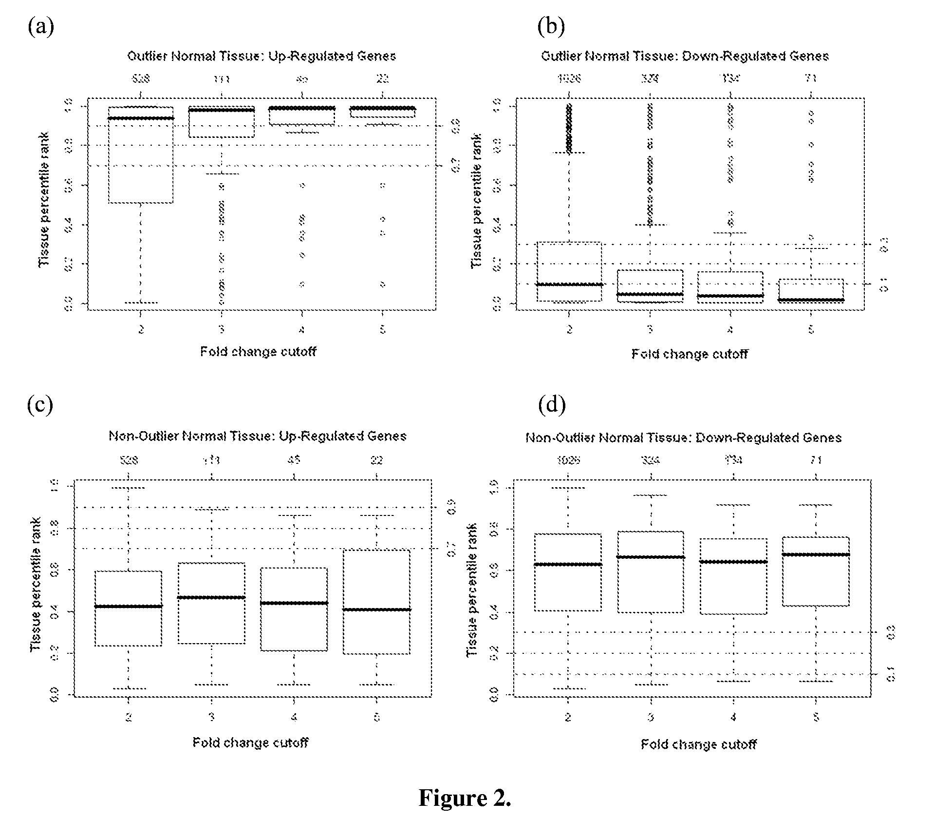
[0152]11 outlier breast tissues were identified using the outlier tissue approach (see, for example, Methods section infra) to re-evaluate the 143 normal breast tissues whose gene expression profiles more closely approximated that of the IDC samples rather than the rest of the 132 normal breast tissues. Eight of these 11 outlier tissues had a median percentile rank greater than 80% among all the 143 normal tissues (i.e., the top 20%) at the 2, 3, and 4 fold-change cutoffs, shown in FIG. 2. The other 3 outlier tissues had a median percentile rank less than 20% (i.e., the bottom 20%) for the under-expressed probe sets. Distinction of the outlier tissues from the normal breast tissues was further demonstrated in FIG. 3.
PUM
| Property | Measurement | Unit |
|---|---|---|
| volume | aaaaa | aaaaa |
| adhesion | aaaaa | aaaaa |
| physical | aaaaa | aaaaa |
Abstract
Description
Claims
Application Information