
Optics and IOLs for Inhibiting cell migration and reduce optic edge dysphotopsia
a technology of optic edge and inhibitory iol, which is applied in the field of intraocular lenses (iols), can solve the problems of reducing image contrast, affecting iol optical performance, and cost of laser treatment, and achieves the effect of inhibiting cell migration and being easy to manufactur
- Summary
- Abstract
- Description
- Claims
- Application Information
AI Technical Summary
Benefits of technology
Problems solved by technology
Method used
Image

Examples
Embodiment Construction
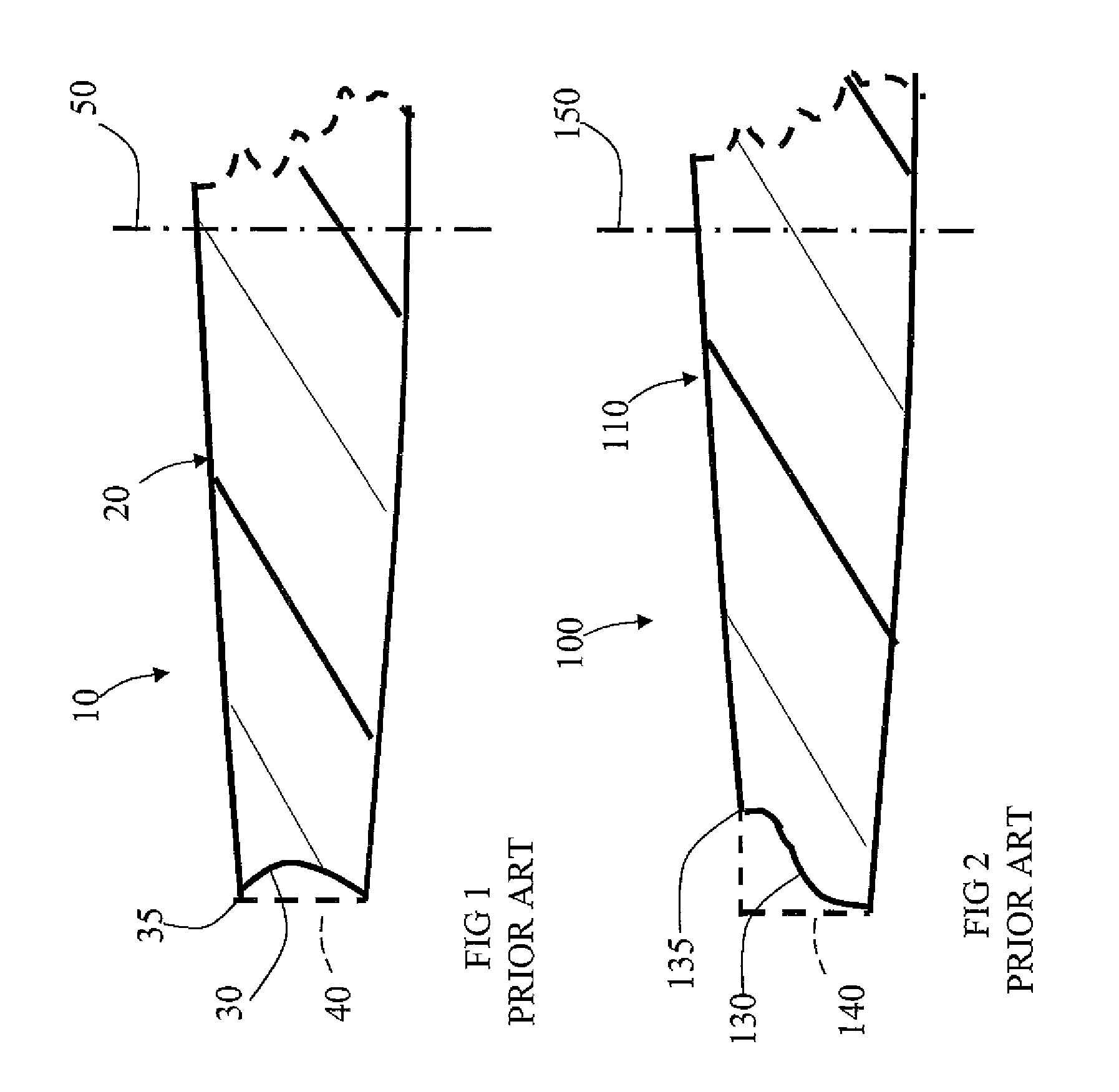
[0022]FIG. 1 illustrates a partial cross-sectional view of an optic 10 of a prior art IOL which has curved optic peripheral edge 30 along the central optical axis 50 as compared with a flat optic peripheral edge 40 (dashed line) that is parallel to the optical axis 50. The optic peripheral edge maintains sharp corner edge 35 with anterior or posterior surface 20 similar to the flat optic peripheral edge 40 to provide inhibition of cell growth. Curved optic peripheral edge 30 provides a reduction in dysphotopsia as compared with flat circular peripheral edge 40 of the optic with substantially constant radius.
[0023]FIG. 2 illustrates a partial cross-sectional view of an optic 100 of another prior art IOL which has more complex curved optic peripheral edge 130 along the central optical axis 150 as compared with the flat optic peripheral edge 140 that is parallel to the optical axis 150. The optic peripheral edge also maintains sharp corner edge 135 with anterior or posterior surface 11...
PUM
 Abstract
Description
Claims
Application Information