Targeting ocular diseases with novel ape1/ref-1 inhibitors

a technology of ape1/ref-1 and inhibitor, which is applied in the field of targeting ocular diseases with novel ape1/ref-1 inhibitors, can solve the problems of possible side effects including hemorrhage and endophthalmitis in patients, and achieve the effects of reducing the ability of hrecs, inhibiting hrec and rf/6a cell migration, and enhancing inhibition of ref-1 function

Active Publication Date: 2021-12-23
INDIANA UNIV RES & TECH CORP
View PDF0 Cites 0 Cited by
  • Summary
  • Abstract
  • Description
  • Claims
  • Application Information

AI Technical Summary

Benefits of technology

[0007]The present disclosure is directed to the use of 3-[(5-(2,3-dimethoxy-6-methyl1,4-benzoquinoyl)]-2-nonyl-2-proprionic acid (APX3330) and / or its derivatives, such [(2E)-2-[(3-methoxy-1,4-dioxo-1,4-dihydronaphthalen-2-yl)methylidene]-N,N-diethylpentanamide] (APX2009) and (2E)-2-[(3-methoxy-1,4-dioxo-1,4-dihydronapthalen-2-yl)methylidene]-N-methoxypentanamide] (APX2014), for inhibiting ocular neovascularization. Particularly, it was found that APX2009 and APX2014 provided enhanced inhibition of Ref-1 function in a DNA-binding assay compared to APX3330. Both compounds were antiproliferative against human retinal microvascular endothelial cells (HRECs; GI50 APX2009: 1.1 μM, APX2014: 110 nM) and macaque choroidal endothelial cells (Rf / 6a GI50 APX2009: 26 μM, APX2014: 5.0 μM). Both compounds significantly reduced the ability of HRECs and Rf / 6a cells to form tubes at mid nanomolar concentrations compared to control, and both significantly inhibited HREC and Rf / 6a cell migration in a scratch wound assay.
[0008]Ex vivo, both APX2009 and APX2014 inhibited choroidal sprouting at low micromolar and high nanomolar concentrations respectively. In the laser-induced choroidal neovascularization mouse model, intraperitoneal APX2009 treatment significantly decreased lesion volume by 4-fold compared to vehicle (p<0.0001, ANOVA with Dunnett's post hoc tests), without obvious intraocular or systemic toxicity. Thus, Ref-1 inhibition with APX2009 and APX2014 blocks ocular angiogenesis in vitro and ex vivo, and APX2009 is an effective systemic therapy for CNV in vivo, establishing Ref-1 inhibition as a promising therapeutic approach for ocular neovascularization.

Problems solved by technology

In all cases, neovessels disrupt retinal architecture and can hemorrhage, leading to blindness.
Although these therapeutic agents have been very successful, significant proportions of patients are resistant and refractory (Lux et al., 2007; Falavarjani and Nguyen, 2013).
Moreover, serious side effects including hemorrhage and endophthalmitis are possible.

Method used


Image

  • Targeting ocular diseases with novel ape1/ref-1 inhibitors
  • Targeting ocular diseases with novel ape1/ref-1 inhibitors
  • Targeting ocular diseases with novel ape1/ref-1 inhibitors

Examples


example 1

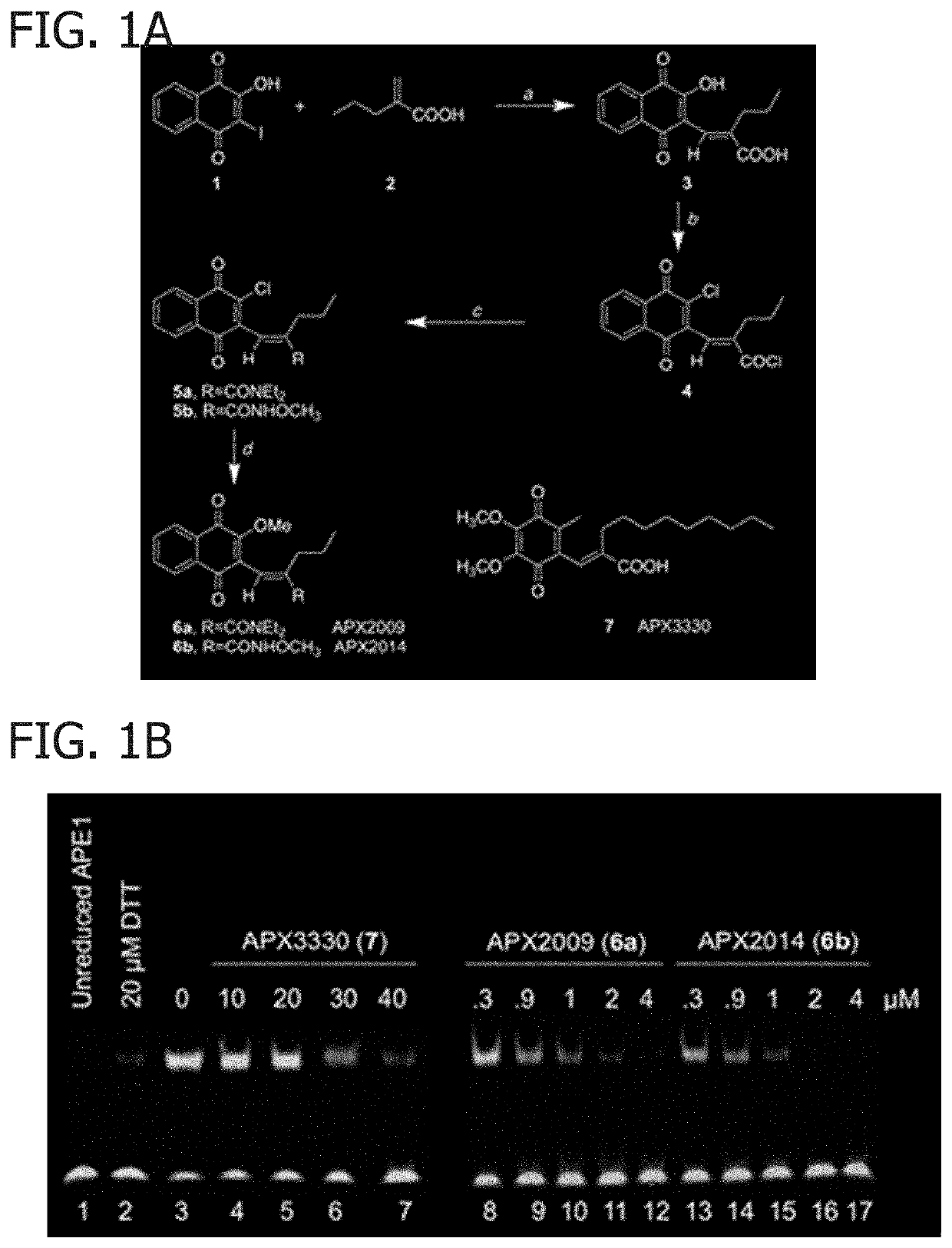
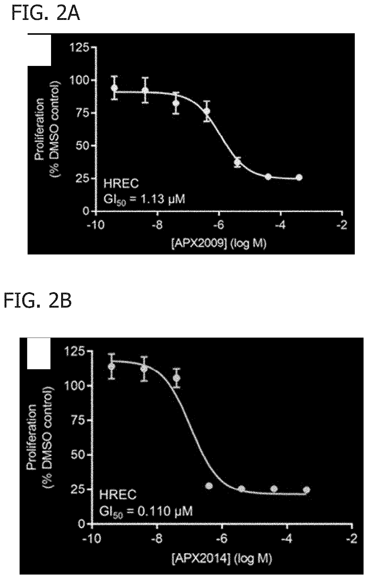
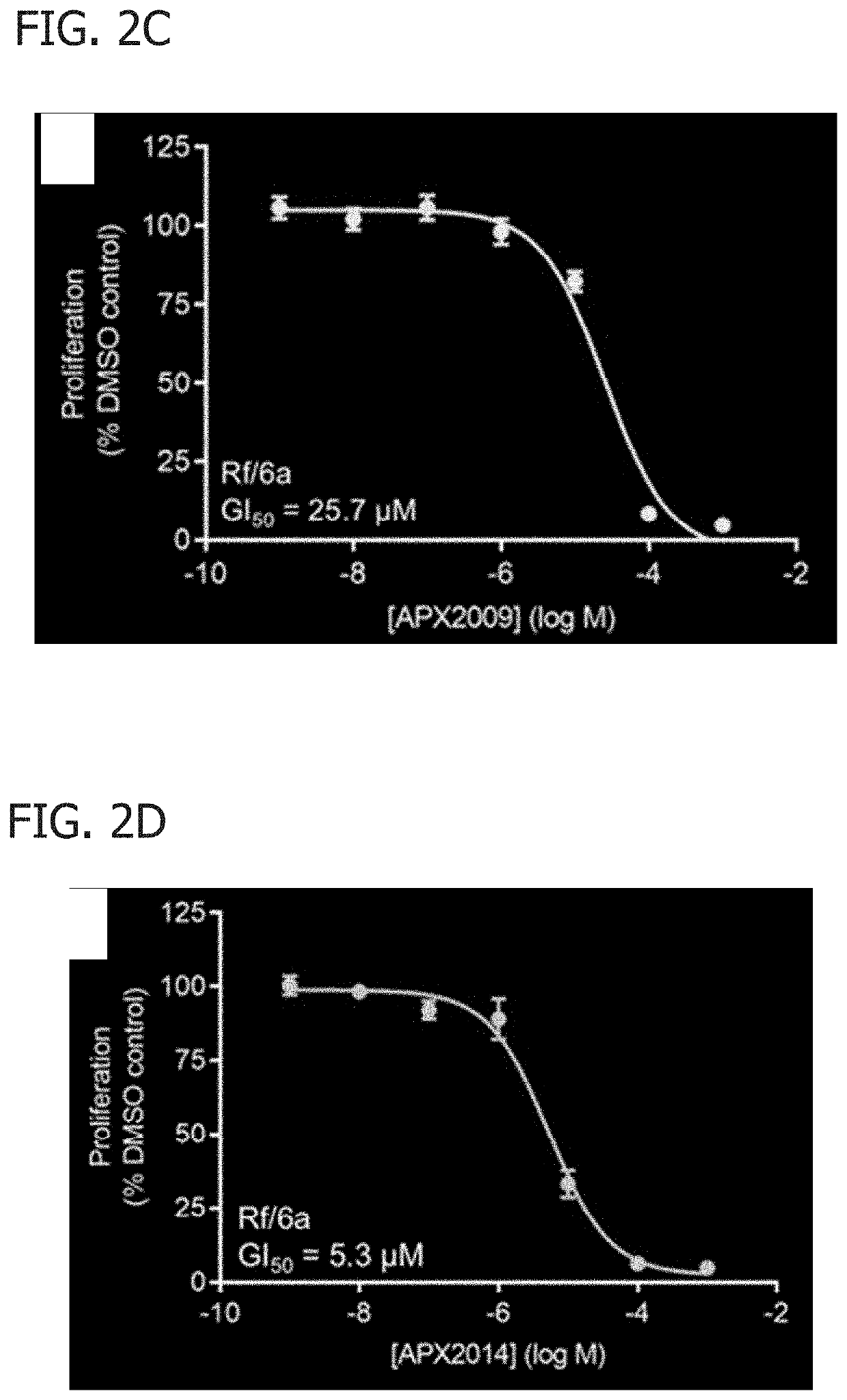
[0048]In this Example, APX2009 and APX2014 were analyzed for their function on AP-1 DNA binding and cell proliferation and migration.

[0049]Materials and Methods

[0050]Synthetic Methods. The compounds were synthesized by Cascade Custom Chemistry (Eugene, Oreg.) and provided by Apexian Pharmaceuticals. In summary (FIG. 1A), iodolawsone (2-iodo-3-hydroxy-1,4 naphthoquinone) was made available from Cascade Custom Chemistry. HPLC were performed using an Alltech Alltima column C18 5u, 250×5 6 mm, flow 1 mL / min at 40° C. Elution was with a mobile phase of 15:10:75 water:A1:methanol where A1 was made using 700 mL of water, 300 mL methanol and 3 mL trimethylamine to which phosphoric acid was added to bring the pH to 3.4.

[0051](E)-2-((3-hydroxy-1,4-dioxo-1,4-dihydronaphthalen-2-yl)methylene)pentanoic acid (3). In a 2 L 3-necked flask equipped with a mechanical stirrer and a gas dispersion fitted tube was placed 2-iodo-3-hydroxy-1,4 naphthoquinone, (iodolawsone, 1) (18 g, 0.06 mol) and 2-propyl...

example 2

[0089]In this Example, the effects of Ref-1 knockdown on NF-κB signaling-associated genes was analyzed.

[0090]Human retinal endothelial cells (HRECs) (Cell Systems, Inc. Kirkland, Wash.) were plated in 6-well plates and treated with 0.1 μM APX2009, 1 μM APX2009 or DMSO for 6 hours and 24 hours. Cells were washed once after treatment with PBS, collected and frozen. This process was repeated to collect treated cells in 4 different passages. RNA was extracted in 300 μL Trizol (Life Technologies, Carlsbad, Calif.) and flash frozen at −80° C. The SMARTer system (Clontech, Mountain View, Calif.) was used to generate cDNA from cells. The dscDNA quantity and quality was assessed using an Agilent Bioanalyzer (Agilent Technologies, Santa Clara, Calif., USA) with the High Sensitivity DNA Chip. A total of 48 SCR and 48 siAPE1 cells were chosen for sequencing. The IUSM Genomics Facility prepared libraries using a Nextera kit (Illumina, San Diego, Calif.). DNAs were sequenced using the Illumina Hi...

example 3

[0092]In this Example, the role of Ref-1 on ocular angiogenesis is analyzed.

[0093]The Protein Atlas was mined for expression data on Ref-1. In addition, immunohistochemistry was performed for Ref-1 in de-identified postmortem eye tissue from a wet AMD patient and an age-matched control. Tissue sections were deparaffinized. DAB was used for detection and counterstained with DAPI. Images of retina and choroid were taken on an EVOS fl digital microscope.

[0094]Ref-1 is highly expressed in developing murine retinas, as well as retinal pigment epithelium (RPE) cells, retinal pericytes, choroidal endothelial cells (CECs) and retinal endothelial cells (RECs). At the RNA level, it is expressed higher in retina than in all but a third of 36 other tissue types (https: / / www.proteinatlas.org / ENSG00000100823-APEX1 / tissue). Preliminary evidence also suggests that it is upregulated in the retina and choroid of human wet AMD patient eyes compared with age-matched controls (FIG. 17), suggesting disea...


PUM

PropertyMeasurementUnit
pHaaaaaaaaaa
pHaaaaaaaaaa
emission wavelengthsaaaaaaaaaa

Abstract

[[(2E)-2-[(3-methoxy-1,4-dioxo-1,4-dihydronaphthalen-2-yl)methylidene]-N,N-diethylpentanamide] (APX2009) and (2E)-2-[(3-methoxy-1,4-dioxo-1,4-dihydronapthalen-2-yl)methylidene]-N-methoxypentanamide] (APX2014) for inhibiting ocular diseases are disclosed herein.

Description

CROSS-REFERENCE TO RELATED APPLICATION[0001]The present disclosure claims priority to U.S. Provisional Application No. 62 / 628,093, filed Feb. 8, 2018, the disclosure of which is hereby incorporated by reference in its entirety.BACKGROUND OF THE DISCLOSURE[0002]The present disclosure relates generally to the use of 3-[(5-(2,3-dimethoxy-6-methyl1,4-benzoquinoyl)]-2-nonyl-2-proprionic acid (APX3330) and / or its derivatives (e.g., [(2E)-2-[(3-methoxy-1,4-dioxo-1,4-dihydro naphthalen-2-yl)methylidene]-N,N-diethylpentanamide] (APX2009), and (2E)-2-[(3-methoxy-1,4-dioxo-1,4-dihydronapthalen-2-yl)methylidene]-N-methoxypentanamide] (APX2014)) for inhibiting ocular diseases.[0003]Ocular neovascularization is the key pathobiological feature of diseases like proliferative diabetic retinopathy (PDR), retinopathy of prematurity (ROP), and wet age-related macular degeneration (AMD), which together are major causes of blindness (Campochiaro, 2013). In PDR and ROP, abnormal blood vessels grow in and ...

Claims


Application Information

Patent Timeline
Patent Type & Authority Applications(United States)
IPC IPC(8): A61K31/165A61K45/06A61P27/02A61K31/675A61K31/45A61K31/445
CPCA61K31/165A61K45/06A61K31/445A61K31/675A61K31/45A61P27/02A61P35/00A61K31/20C07C66/00A61K2300/00A61K31/122C07C50/06C07C50/12
Inventor KELLEY, MARK R.CORSON, TIMOTHY W.
Owner INDIANA UNIV RES & TECH CORP
Why Eureka
Try Eureka