Method and System for Computational Modeling of the Aorta and Heart

a computational modeling and aorta technology, applied in the field of medical image modeling, can solve the problems of ineffective therapeutic intervention for individual patients, lack of accurate estimates, and large incidence of premature morbidity and mortality

Active Publication Date: 2011-03-10
SIEMENS HEALTHCARE GMBH
View PDF10 Cites 162 Cited by
  • Summary
  • Abstract
  • Description
  • Claims
  • Application Information

AI Technical Summary

Benefits of technology

[0006]In one embodiment of the present invention, a multi-component patient specific 4D geometric model of the heart and aorta is estimated from a sequence of volumetric cardiac imaging data. A patient specific 4D computational model is generated based on one or more of personalized geometry, material properties, fluid boundary conditions, and flow velocity measurements in the 4D geometric model. Patient specific material properties of the aortic wall are estimated based on the 4D geometrical model and the 4D computational model. A patient specific biomechanical model can be generated based on Fluid Structure Interaction (FSI) simulations using the 4D computational model and the estimated material properties of the aortic wall. Patient specific clinical parameters can be extracted based on the 4D geometric model and the FSI simulations. Disease progression modeling and risk stratification can be performed based on the patient specific clinical parameters.

Problems solved by technology

Although medical advances in recent years have provided important improvements in the diagnosis and treatment of complex cardiac diseases such as valvular disease, thoracic aortic aneurysm, and Tetralogy of Fallot, the incidence of premature morbidity and mortality is still large.
These problems are due, at least in part, to a lack of accurate estimates (in-vivo and in-vitro) of patient-specific parameters that accurately characterize the heart and aortic anatomy, physiology and hemodynamics.
As a result, early disease prediction and progression models are often based on generic data, rendering them ineffective for therapeutic interventions on individual patients.
However, there has been little focus on extending the hemodynamic analysis by incorporating patient-specific geometry, boundary conditions, and material properties for the vascular structures.

Method used


Image

  • Method and System for Computational Modeling of the Aorta and Heart
  • Method and System for Computational Modeling of the Aorta and Heart
  • Method and System for Computational Modeling of the Aorta and Heart

Examples


Embodiment Construction

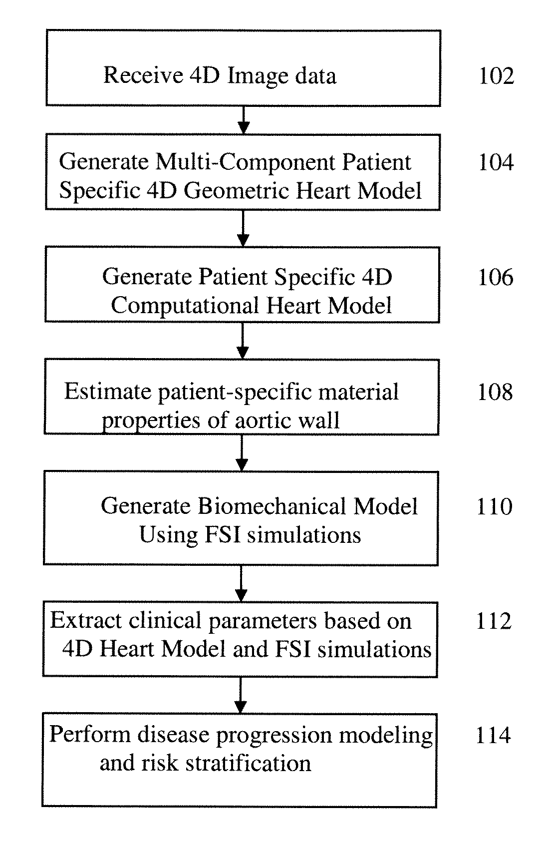
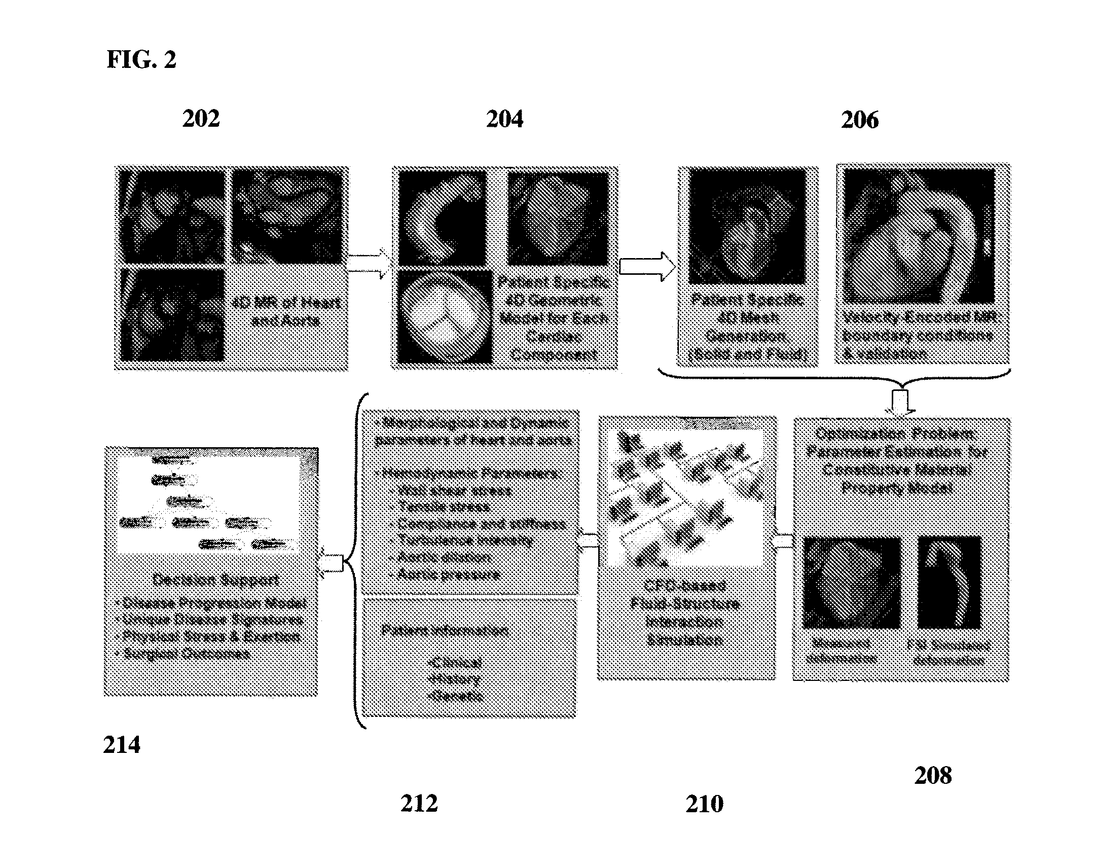
[0020]The present invention relates to generating a 4D personalized anatomical model of the heart from a sequence of volumetric data, such as computed tomography (CT), magnetic resonance imaging (MRI), and echocardiography data. Such sequences of volumetric data also referred to herein as 4D image data or 4D images, are sequences taken over a period of time to cover one or more cardiac cycles, in which each frame is a 3D image (volume). Embodiments of the present invention are described herein to give a visual understanding of the heart modeling method. A digital image is often composed of digital representations of one or more objects (or shapes). The digital representation of an object is often described herein in terms of identifying and manipulating the objects. Such manipulations are virtual manipulations accomplished in the memory or other circuitry / hardware of a computer system. Accordingly, is to be understood that embodiments of the present invention may be performed within...


PUM


Abstract

A method and system for generating a patient specific anatomical heart model is disclosed. A sequence of volumetric image data, such as computed tomography (CT), echocardiography, or magnetic resonance (MR) image data of a patient's cardiac region is received. A multi-component patient specific 4D geometric model of the heart and aorta estimated from the sequence of volumetric cardiac imaging data. A patient specific 4D computational model based on one or more of personalized geometry, material properties, fluid boundary conditions, and flow velocity measurements in the 4D geometric model is generated. Patient specific material properties of the aortic wall are estimated using the 4D geometrical model and the 4D computational model. Fluid Structure Interaction (FSI) simulations are performed using the 4D computational model and estimated material properties of the aortic wall, and patient specific clinical parameters are extracted based on the FSI simulations. Disease progression modeling and risk stratification are performed based on the patient specific clinical parameters.

Description

[0001]This application claims the benefit of U.S. Provisional Application No. 61 / 240,397, filed Sep. 8, 2009, the disclosure of which is herein incorporated by reference.BACKGROUND OF THE INVENTION[0002]The present invention relates to modeling the heart in medical images, and more particularly, to modeling the heart using personalized 4D anatomical heart model of the full cardiac system estimated from volumetric image sequences for decision support in diagnosis and treatment of cardiac disease.[0003]Cardiac disease is the leading cause of death for men and women in the United States and accounts no less than 30% of deaths worldwide. Although medical advances in recent years have provided important improvements in the diagnosis and treatment of complex cardiac diseases such as valvular disease, thoracic aortic aneurysm, and Tetralogy of Fallot, the incidence of premature morbidity and mortality is still large. These problems are due, at least in part, to a lack of accurate estimates...

Claims


Application Information

Patent Timeline
Patent Type & Authority Applications(United States)
IPC IPC(8): G06G7/60
CPCG06F19/3437G06F19/345G06T2207/30048G06T17/00G06T2207/10072G06T7/0012G16H50/50G16H50/20
Inventor SHARMA, PUNEETGEORGESCU, BOGDANIONASEC, RAZVAN IOANCOMANICIU, DORIN
Owner SIEMENS HEALTHCARE GMBH
Why Eureka
Try Eureka